NP234690A94

c-Myb Antibody (MYB286), Alexa Fluor™ 594, Novus Biologicals™

Manufacturer: Novus Biologicals

Select a Size

Pack Size SKU Availability Price
Each of 1 NP234690A94-Each-of-1 In Stock ₹ 55,271.76

NP234690A94 - Each of 1

₹ 55,271.76

In Stock

Quantity

1

Base Price: ₹ 55,271.76

GST (18%): ₹ 9,948.917

Total Price: ₹ 65,220.677

Antigen

c-Myb

Classification

Monoclonal

Conjugate

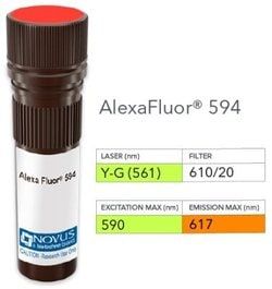
Alexa Fluor 594

Formulation

50 mM sodium borate with 0.05% sodium azide

Gene Symbols

MYB

Immunogen

A synthetic peptide, corresponding to aa 119-135 (RRKVEQEGYPQESSKAG) of c-myb onco protein, coupled to KLH (Uniprot: P01104)

Quantity

0.1 mL

Gene ID (Entrez)

4602

Target Species

Human

Isotype

IgG1 κ

Applications

Western Blot, Flow Cytometry, ELISA, Immunocytochemistry, Immunofluorescence

Clone

MYB286

Dilution

Western Blot, Flow Cytometry, ELISA, Immunocytochemistry/Immunofluorescence

Gene Alias

c-myb, Cmyb, c-myb protein (140 AA), c-myb_CDS, c-myb10A_CDS, c-myb13A_CDS, c-myb14A_CDS, c-myb8B_CDS, efg, Proto-oncogene c-Myb, transcriptional activator Myb, v-myb avian myeloblastosis viral oncogene homolog, v-myb myeloblastosis viral oncogene homolog (avian)

Host Species

Mouse

Purification Method

Protein A or G purified

Primary or Secondary

Primary

Test Specificity

The highly leukemogenic avian retrovirus E26 contains two oncogenes, v-Myb and v-Ets, which are expressed together as a fusion protein. The cellular homolog of v-Myb, designated c-Myb, encodes a transcription factor. Deletion or disruption of a negative regulatory domain mapping within the carboxy-terminal domain of c-Myb results in enhanced transactivating capacity and in parallel, leads to activation of its ability to transform hemopoietic cells. c-Myb is expressed preferentially, but not exclusively, in immature hemopoietic cells and its expression decreases as cells differentiate.

Content And Storage

Store at 4°C in the dark.

Related Products

Img

Novus Biologicals

NB600302FR

--

Img

Novus Biologicals

NB303844

--

Img

Novus Biologicals

NB247739488

--

Img

Novus Biologicals

NP233313A32

--

Img

Novus Biologicals

NP23413A94

--

Img

Novus Biologicals

NP234646A70

--

Img

Novus Biologicals

NB239856

--

Img

Novus Biologicals

NBP177323

--

Description

  • c-Myb Monoclonal specifically detects c-Myb in Human samples
  • It is validated for ELISA.

Compare Similar Items

Show Difference

Img

Novus Biologicals

NP234690A94

--


Antigen:
c-Myb

Classification:
Monoclonal

Conjugate:
Alexa Fluor 594

Formulation:
50 mM sodium borate with 0.05% sodium azide

Gene Symbols:
MYB

Immunogen:
A synthetic peptide, corresponding to aa 119-135 (RRKVEQEGYPQESSKAG) of c-myb onco protein, coupled to KLH (Uniprot: P01104)

Quantity:
0.1 mL

Gene ID (Entrez):
4602

Target Species:
Human

Isotype:
IgG1 κ

Applications:
Western Blot, Flow Cytometry, ELISA, Immunocytochemistry, Immunofluorescence

Clone:
MYB286

Dilution:
Western Blot, Flow Cytometry, ELISA, Immunocytochemistry/Immunofluorescence

Gene Alias:
c-myb, Cmyb, c-myb protein (140 AA), c-myb_CDS, c-myb10A_CDS, c-myb13A_CDS, c-myb14A_CDS, c-myb8B_CDS, efg, Proto-oncogene c-Myb, transcriptional activator Myb, v-myb avian myeloblastosis viral oncogene homolog, v-myb myeloblastosis viral oncogene homolog (avian)

Host Species:
Mouse

Purification Method:
Protein A or G purified

Primary or Secondary:
Primary

Test Specificity:
The highly leukemogenic avian retrovirus E26 contains two oncogenes, v-Myb and v-Ets, which are expressed together as a fusion protein. The cellular homolog of v-Myb, designated c-Myb, encodes a transcription factor. Deletion or disruption of a negative regulatory domain mapping within the carboxy-terminal domain of c-Myb results in enhanced transactivating capacity and in parallel, leads to activation of its ability to transform hemopoietic cells. c-Myb is expressed preferentially, but not exclusively, in immature hemopoietic cells and its expression decreases as cells differentiate.

Content And Storage:
Store at 4°C in the dark.

Img

Novus Biologicals

NP234691A30

--


Antigen:
c-Myc

Classification:
Monoclonal

Conjugate:
Alexa Fluor 350

Formulation:
50 mM sodium borate with 0.05% sodium azide

Gene Symbols:
MYC

Immunogen:
Recombinant human c-Myc protein (Uniprot: P01106)

Quantity:
0.1 mL

Gene ID (Entrez):
4609

Target Species:
Human

Isotype:
IgG1 κ

Applications:
Flow Cytometry, Immunohistochemistry, Immunocytochemistry, Immunofluorescence, Immunohistochemistry (Paraffin)

Clone:
MYC275

Dilution:
Flow Cytometry, Immunohistochemistry, Immunocytochemistry/Immunofluorescence, Immunohistochemistry-Paraffin

Gene Alias:
avian myelocytomatosis viral oncogene homolog, BHLHE39, bHLHe39MRTL, Class E basic helix-loop-helix protein 39, c-Myc, MYC, myc proto-oncogene protein, MYCC, myc-related translation/localization regulatory factor, Proto-oncogene c-Myc, Transcription factor p64, v-myc avian myelocytomatosis viral oncogene homolog, v-myc myelocytomatosis viral oncogene homolog (avian)

Host Species:
Mouse

Purification Method:
__

Primary or Secondary:
Primary

Test Specificity:
The c-Myc protein is a transcription factor, which is encoded by the c-Myc gene on human chromosome 8q24. c-Myc is commonly activated in a variety of tumor cells and plays an important role in cellular proliferation, differentiation, apoptosis and cell cycle progression. The phosphorylation of c-Myc has been investigated and previous studies have suggested a functional association between phosphorylation at Thr58/Ser62 by glycogen synthase kinase 3, cyclin dependent kinase, ERK2 and C-Jun N terminal Kinase (JNK) in cell proliferation and cell cycle regulation. Studies also have shown that c-Myc is essential for tumor cell development in vasculogenesis and angiogenesis that distribute blood throughout the cells, and which brought extensive attention in the development of new therapeutic approach for cancer treatment.

Content And Storage:
Store at 4°C in the dark.

Img

Novus Biologicals

NP234691A32

--


Antigen:
c-Myc

Classification:
Monoclonal

Conjugate:
Alexa Fluor 532

Formulation:
50 mM sodium borate with 0.05% sodium azide

Gene Symbols:
MYC

Immunogen:
Recombinant human c-Myc protein (Uniprot: P01106)

Quantity:
0.1 mL

Gene ID (Entrez):
4609

Target Species:
Human

Isotype:
IgG1 κ

Applications:
Flow Cytometry, Immunohistochemistry, Immunocytochemistry, Immunofluorescence, Immunohistochemistry (Paraffin)

Clone:
MYC275

Dilution:
Flow Cytometry, Immunohistochemistry, Immunocytochemistry/Immunofluorescence, Immunohistochemistry-Paraffin

Gene Alias:
avian myelocytomatosis viral oncogene homolog, BHLHE39, bHLHe39MRTL, Class E basic helix-loop-helix protein 39, c-Myc, MYC, myc proto-oncogene protein, MYCC, myc-related translation/localization regulatory factor, Proto-oncogene c-Myc, Transcription factor p64, v-myc avian myelocytomatosis viral oncogene homolog, v-myc myelocytomatosis viral oncogene homolog (avian)

Host Species:
Mouse

Purification Method:
__

Primary or Secondary:
Primary

Test Specificity:
The c-Myc protein is a transcription factor, which is encoded by the c-Myc gene on human chromosome 8q24. c-Myc is commonly activated in a variety of tumor cells and plays an important role in cellular proliferation, differentiation, apoptosis and cell cycle progression. The phosphorylation of c-Myc has been investigated and previous studies have suggested a functional association between phosphorylation at Thr58/Ser62 by glycogen synthase kinase 3, cyclin dependent kinase, ERK2 and C-Jun N terminal Kinase (JNK) in cell proliferation and cell cycle regulation. Studies also have shown that c-Myc is essential for tumor cell development in vasculogenesis and angiogenesis that distribute blood throughout the cells, and which brought extensive attention in the development of new therapeutic approach for cancer treatment.

Content And Storage:
Store at 4°C in the dark.

Img

Novus Biologicals

NP234694A30

--


Antigen:
CD63

Classification:
Monoclonal

Conjugate:
Alexa Fluor 350

Formulation:
50 mM sodium borate with 0.05% sodium azide

Gene Symbols:
CD63

Immunogen:
Smooth plasma membrane fraction of MeWo cells

Quantity:
0.1 mL

Gene ID (Entrez):
967

Target Species:
Human, Mouse

Isotype:
IgG1 κ

Applications:
Western Blot, Flow Cytometry, ELISA, Immunohistochemistry, Immunocytochemistry, Immunofluorescence, Immunohistochemistry (Paraffin)

Clone:
NKI/C3

Dilution:
Western Blot, Flow Cytometry, ELISA, Immunohistochemistry, Immunocytochemistry/Immunofluorescence, Immunohistochemistry-Paraffin, Immunohistochemistry-Frozen

Gene Alias:
CD63 antigen, CD63 antigen (melanoma 1 antigen), CD63 molecule, Granulophysin, LAMP-3, Lysosomal-associated membrane protein 3, ME491, melanoma 1 antigen, Melanoma-associated antigen ME491, melanoma-associated antigen MLA1, MLA1lysosome-associated membrane glycoprotein 3, Ocular melanoma-associated antigen, OMA81H, Tetraspanin-30, tspan-30, TSPAN30granulophysin

Host Species:
Mouse

Purification Method:
__

Primary or Secondary:
Primary

Test Specificity:
This monoclonal antibody recognizes protein of 26kDa-60kDa, which is identified as CD63. Its epitope is different from that of monoclonal antibody LAMP3/529. The tetraspanins are integral membrane proteins expressed on cell surface and granular membranes of hematopoietic cells and are components of multi-molecular complexes with specific integrins. The tetraspanin CD63 is a lysosomal membrane glycoprotein that translocates to the plasma membrane after platelet activation. CD63 is expressed on activated platelets, monocytes and macrophages, and is weakly expressed on granulocytes, T cell and B cells. It is located on the basophilic granule membranes and on the plasma membranes of lymphocytes and granulocytes. CD63 is a member of the TM4 superfamily of leukocyte glycoproteins that includes CD9, CD37 and CD53, which contain four transmembrane regions. CD63 may play a role in phagocytic and intracellular lysosome-phagosome fusion events. CD63 deficiency is associated with Hermansky-Pudlak

Content And Storage:
Store at 4°C in the dark.