Nuclear Membrane Marker Antibody (NM97), Alexa Fluor™ 594, Novus Biologicals™
Manufacturer: Novus Biologicals
Select a Size
| Pack Size | SKU | Availability | Price |
|---|---|---|---|
| Each of 1 | NP234696A94-Each-of-1 | In Stock | ₹ 57,494.00 |
NP234696A94 - Each of 1
In Stock
Quantity
1
Base Price: ₹ 57,494.00
GST (18%): ₹ 10,348.92
Total Price: ₹ 67,842.92
Antigen
Nuclear Membrane Marker
Classification
Monoclonal
Conjugate
Alexa Fluor 594
Formulation
50 mM sodium borate with 0.05% sodium azide
Immunogen
Nuclei of myeloid leukemia biopsy cells
Quantity
0.1 mL
Test Specificity
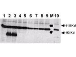
This monoclonal antibody is part of a new panel of reagents, which recognizes subcellular organelles or compartments of human cells. These markers may be useful in identification of these organelles in cells, tissues, and biochemical preparations. It recognizes an antigen associated with the nuclear membrane expressed in human cells. It can be used to stain the nuclear membrane in cell or tissue preparations and can be used as a marker of the nuclear membrane in subcellular fractions. It produces a ring pattern around the nucleus of cells of normal and malignant cells and may be used to stain the nuclear membrane of cells in fixed or frozen tissue sections. The nuclear envelope (also known as the perinuclear envelope, nuclear membrane, nucleolemma or karyotheca) is the double membrane of the nucleus that encloses genetic material in eukaryotic cells. It separates the contents of the nucleus (DNA in particular) from the cytosol (cytoplasm). Numerous nuclear pores are present on the nucl
Content And Storage
Store at 4°C in the dark.
Applications
Immunocytochemistry, Immunofluorescence
Clone
NM97
Dilution
Immunocytochemistry/Immunofluorescence
Host Species
Mouse
Purification Method
Protein A or G purified
Primary or Secondary
Primary
Target Species
Human
Isotype
IgG1 κ
Related Products

Description
- Nuclear Membrane Marker Monoclonal specifically detects Nuclear Membrane Marker in Human samples
- It is validated for Immunocytochemistry/Immunofluorescence.
Compare Similar Items
Show Difference
Antigen: Nuclear Membrane Marker
Classification: Monoclonal
Conjugate: Alexa Fluor 594
Formulation: 50 mM sodium borate with 0.05% sodium azide
Immunogen: Nuclei of myeloid leukemia biopsy cells
Quantity: 0.1 mL
Test Specificity: This monoclonal antibody is part of a new panel of reagents, which recognizes subcellular organelles or compartments of human cells. These markers may be useful in identification of these organelles in cells, tissues, and biochemical preparations. It recognizes an antigen associated with the nuclear membrane expressed in human cells. It can be used to stain the nuclear membrane in cell or tissue preparations and can be used as a marker of the nuclear membrane in subcellular fractions. It produces a ring pattern around the nucleus of cells of normal and malignant cells and may be used to stain the nuclear membrane of cells in fixed or frozen tissue sections. The nuclear envelope (also known as the perinuclear envelope, nuclear membrane, nucleolemma or karyotheca) is the double membrane of the nucleus that encloses genetic material in eukaryotic cells. It separates the contents of the nucleus (DNA in particular) from the cytosol (cytoplasm). Numerous nuclear pores are present on the nucl
Content And Storage: Store at 4°C in the dark.
Applications: Immunocytochemistry, Immunofluorescence
Clone: NM97
Dilution: Immunocytochemistry/Immunofluorescence
Host Species: Mouse
Purification Method: Protein A or G purified
Primary or Secondary: Primary
Target Species: Human
Isotype: IgG1 κ
Antigen: CD1a
Classification: Monoclonal
Conjugate: Alexa Fluor 350
Formulation: 50 mM sodium borate with 0.05% sodium azide
Immunogen: Human thymus cells
Quantity: 0.1 mL
Test Specificity: At least five CD1 genes (CD1a, b, c, d, and e) are identified. CD1 proteins have been demonstrated to restrict T cell response to non-peptide lipid and glycolipid antigens and play a role in non-classical antigen presentation. CD1a is a non-polymorphic MHC Class 1 related cell surface glycoprotein, expressed in association with Beta-2 microglobulin. Anti-CD1a labels Langerhans cell histiocytosis (Histiocytosis X), extranodal histiocytic sarcoma, a subset of T-lymphoblastic lymphoma/leukemia, and interdigitating dendritic cell sarcoma of the lymph node. When combined with antibodies against TTF-1 and CD5, anti-CD1a is useful in distinguishing between pulmonary and thymic neoplasms since CD1a is consistently expressed in thymic lymphocytes in both typical and atypical thymomas, but only focally in 1/6 of thymic carcinomas and not in lymphocytes in pulmonary neoplasms. Anti-CD1a is reported to be a new marker for perivascular epithelial cell tumor (PEComa).
Content And Storage: Store at 4°C in the dark.
Applications: Western Blot, Flow Cytometry, ELISA, Immunohistochemistry, Immunocytochemistry, Immunofluorescence, Immunohistochemistry (Paraffin)
Clone: O10
Dilution: Western Blot, Flow Cytometry, ELISA, Immunohistochemistry, Immunocytochemistry/Immunofluorescence, Immunohistochemistry-Paraffin, Immunohistochemistry-Frozen
Host Species: Mouse
Purification Method: Protein A or G purified
Primary or Secondary: Primary
Target Species: Human
Isotype: IgG1 κ
Antigen: CD1a
Classification: Monoclonal
Conjugate: Alexa Fluor 532
Formulation: 50 mM sodium borate with 0.05% sodium azide
Immunogen: Human thymus cells
Quantity: 0.1 mL
Test Specificity: At least five CD1 genes (CD1a, b, c, d, and e) are identified. CD1 proteins have been demonstrated to restrict T cell response to non-peptide lipid and glycolipid antigens and play a role in non-classical antigen presentation. CD1a is a non-polymorphic MHC Class 1 related cell surface glycoprotein, expressed in association with Beta-2 microglobulin. Anti-CD1a labels Langerhans cell histiocytosis (Histiocytosis X), extranodal histiocytic sarcoma, a subset of T-lymphoblastic lymphoma/leukemia, and interdigitating dendritic cell sarcoma of the lymph node. When combined with antibodies against TTF-1 and CD5, anti-CD1a is useful in distinguishing between pulmonary and thymic neoplasms since CD1a is consistently expressed in thymic lymphocytes in both typical and atypical thymomas, but only focally in 1/6 of thymic carcinomas and not in lymphocytes in pulmonary neoplasms. Anti-CD1a is reported to be a new marker for perivascular epithelial cell tumor (PEComa).
Content And Storage: Store at 4°C in the dark.
Applications: Western Blot, Flow Cytometry, ELISA, Immunohistochemistry, Immunocytochemistry, Immunofluorescence, Immunohistochemistry (Paraffin)
Clone: O10
Dilution: Western Blot, Flow Cytometry, ELISA, Immunohistochemistry, Immunocytochemistry/Immunofluorescence, Immunohistochemistry-Paraffin, Immunohistochemistry-Frozen
Host Species: Mouse
Purification Method: Protein A or G purified
Primary or Secondary: Primary
Target Species: Human
Isotype: IgG1 κ