PLC/PRF/5 (human hepatoma) Nuclear extract lysate, Non-denatured; Abnova
Manufacturer: Abnova Corporation
Select a Size
| Pack Size | SKU | Availability | Price |
|---|---|---|---|
| Each of 1 | 89-014-445-Each-of-1 | In Stock | ₹ 42,720.00 |
89-014-445 - Each of 1
In Stock
Quantity
1
Base Price: ₹ 42,720.00
GST (18%): ₹ 7,689.60
Total Price: ₹ 50,409.60
Tissue
Liver
Description
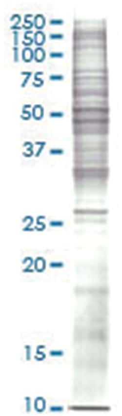
Nuclear extract cell lysate (non-denatured)
Preparation Method
Nuclear extract was prepared by using a modified protocol of Dignam et al. Cells were Harvested and homogenized in Buffer A, and then centrifugated at 25,000 g for 20 minutes to remove cytoplasm and pellet the nuclei. The pellet was re-suspended in Buffer C, and then the suspensions were centrifuged to collect nuclear extract. The supernatant was dialyzed against Buffer D. The dialysate was then centrifuged, divided into aliquots, and stored at -80°C. The protein concentration was determined by the method of Bradford (Bio-Rad protein assay, microplate standard assay). The lysate was adjusted to 2 mg/ml.
Quantity
50 μg
Host Species
Human
Content And Storage
Store at -80°C. Aliquot to avoid repeated freezing and thawing.
For Use With (Application)
Immunoprecipitation, Western Blot
Lysis Buffer
Buffer A: 10mM HEPES pH 7.9, 1.5mM MgCl2, 10mM KCl, 0.5 mM DTT. Buffer C: 20mM HEPES pH 7.9, 25%(v/v) Glycerol , 0.42M NaCl , 1.5mM MgCl2, 0.2 mM EDTA, 0.5 mM DTT & 0.5 mM PMSF. Buffer D : 20mM HEPES pH 7.9, 20%(v/v) glycerol, 50mM KCl, 0.2 mM EDTA, 0.5 mM DTT & 0.5 mM PMSF.
Quality Control Testing
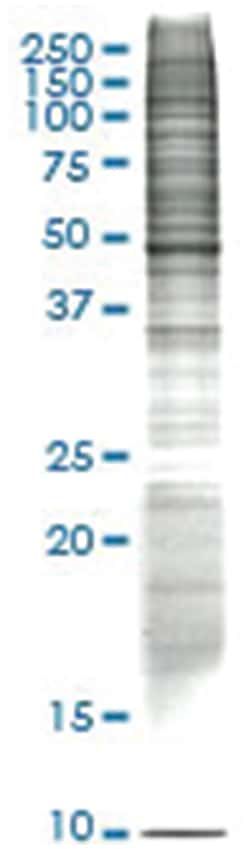
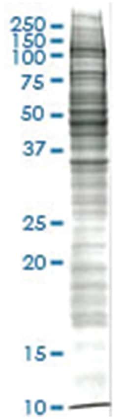
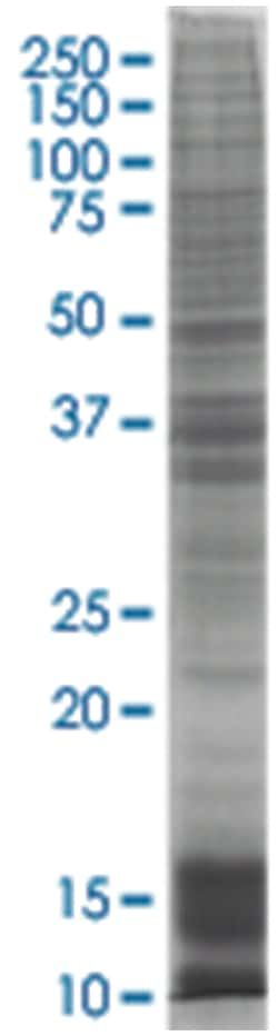
12.5% SDS-PAGE Stained with Coomassie Blue.
Storage Buffer
In Buffer D.
Concentration
2.5 mg/mL
Related Products

Description
- Western Blotting, Immunoprecipitation
Compare Similar Items
Show Difference
Tissue: Liver
Description: Nuclear extract cell lysate (non-denatured)
Preparation Method: Nuclear extract was prepared by using a modified protocol of Dignam et al. Cells were Harvested and homogenized in Buffer A, and then centrifugated at 25,000 g for 20 minutes to remove cytoplasm and pellet the nuclei. The pellet was re-suspended in Buffer C, and then the suspensions were centrifuged to collect nuclear extract. The supernatant was dialyzed against Buffer D. The dialysate was then centrifuged, divided into aliquots, and stored at -80°C. The protein concentration was determined by the method of Bradford (Bio-Rad protein assay, microplate standard assay). The lysate was adjusted to 2 mg/ml.
Quantity: 50 μg
Host Species: Human
Content And Storage: Store at -80°C. Aliquot to avoid repeated freezing and thawing.
For Use With (Application): Immunoprecipitation, Western Blot
Lysis Buffer: Buffer A: 10mM HEPES pH 7.9, 1.5mM MgCl2, 10mM KCl, 0.5 mM DTT. Buffer C: 20mM HEPES pH 7.9, 25%(v/v) Glycerol , 0.42M NaCl , 1.5mM MgCl2, 0.2 mM EDTA, 0.5 mM DTT & 0.5 mM PMSF. Buffer D : 20mM HEPES pH 7.9, 20%(v/v) glycerol, 50mM KCl, 0.2 mM EDTA, 0.5 mM DTT & 0.5 mM PMSF.
Quality Control Testing: 12.5% SDS-PAGE Stained with Coomassie Blue.
Storage Buffer: In Buffer D.
Concentration: 2.5 mg/mL
Tissue: Adrenal Gland
Description: Nuclear extract cell lysate (non-denatured)
Preparation Method: Nuclear extract was prepared by using a modified protocol of Dignam et al. Cells were Harvested and homogenized in Buffer A, and then centrifugated at 25,000 g for 20 minutes to remove cytoplasm and pellet the nuclei. The pellet was re-suspended in Buffer C, and then the suspensions were centrifuged to collect nuclear extract. The supernatant was dialyzed against Buffer D. The dialysate was then centrifuged, divided into aliquots, and stored at -80°C. The protein concentration was determined by the method of Bradford (Bio-Rad protein assay, microplate standard assay). The lysate was adjusted to 2 mg/ml.
Quantity: 50 μg
Host Species: Rat
Content And Storage: Store at -80°C. Aliquot to avoid repeated freezing and thawing.
For Use With (Application): Immunoprecipitation, Western Blot
Lysis Buffer: Buffer A: 10mM HEPES pH 7.9, 1.5mM MgCl2, 10mM KCl, 0.5 mM DTT. Buffer C: 20mM HEPES pH 7.9, 25%(v/v) Glycerol , 0.42M NaCl , 1.5mM MgCl2, 0.2 mM EDTA, 0.5 mM DTT & 0.5 mM PMSF. Buffer D : 20mM HEPES pH 7.9, 20%(v/v) glycerol, 50mM KCl, 0.2 mM EDTA, 0.5 mM DTT & 0.5 mM PMSF.
Quality Control Testing: 12.5% SDS-PAGE Stained with Coomassie Blue.
Storage Buffer: In Buffer D.
Concentration: 2.5 mg/mL
Tissue: Blood, peripheral
Description: Nuclear extract cell lysate (non-denatured)
Preparation Method: Nuclear extract was prepared by using a modified protocol of Dignam et al. Cells were Harvested and homogenized in Buffer A, and then centrifugated at 25,000 g for 20 minutes to remove cytoplasm and pellet the nuclei. The pellet was re-suspended in Buffer C, and then the suspensions were centrifuged to collect nuclear extract. The supernatant was dialyzed against Buffer D. The dialysate was then centrifuged, divided into aliquots, and stored at -80°C. The protein concentration was determined by the method of Bradford (Bio-Rad protein assay, microplate standard assay). The lysate was adjusted to 2 mg/ml.
Quantity: 50 μg
Host Species: Human
Content And Storage: Store at -80°C. Aliquot to avoid repeated freezing and thawing.
For Use With (Application): Immunoprecipitation, Western Blot
Lysis Buffer: Buffer A: 10mM HEPES pH 7.9, 1.5mM MgCl2, 10mM KCl, 0.5 mM DTT. Buffer C: 20mM HEPES pH 7.9, 25%(v/v) Glycerol , 0.42M NaCl , 1.5mM MgCl2, 0.2 mM EDTA, 0.5 mM DTT & 0.5 mM PMSF. Buffer D : 20mM HEPES pH 7.9, 20%(v/v) glycerol, 50mM KCl, 0.2 mM EDTA, 0.5 mM DTT & 0.5 mM PMSF.
Quality Control Testing: 12.5% SDS-PAGE Stained with Coomassie Blue.
Storage Buffer: In Buffer D.
Concentration: 2.5 mg/mL