MABS2282
Anti-SIRPA Antibody, clone MY-1
Manufacturer: Sigma Aldrich
Synonym(S): Tyrosine-protein phosphatase non-receptor type substrate 1;SHP substrate 1;SHPS-1;Brain Ig-like molecule with tyrosine-based activation motifs;Bit;CD172 antigen-like family member A;Inhibitory receptor SHPS-1;MyD-1 antigen;Signal-regulatory protein alpha-
Select a Size
| Pack Size | SKU | Availability | Price |
|---|---|---|---|
| 25 μL | MABS2282-25-μL | In Stock | ₹ 13,779.99 |
| 100 μL | MABS2282-100-μL | In Stock | ₹ 34,740.00 |
MABS2282 - 25 μL
In Stock
Quantity
1
Base Price: ₹ 13,779.99
GST (18%): ₹ 2,480.398
Total Price: ₹ 16,260.388
biological source
rat
Quality Level
200
conjugate
unconjugated
antibody form
purified antibody
antibody product type
primary antibodies
clone
MY-1, monoclonal
mol wt
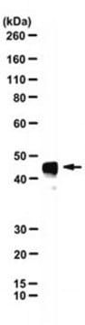
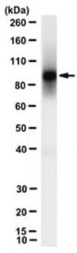
calculated mol wt 56.14 kDaobserved mol wt ~95 kDa
purified by
using protein G
species reactivity
mouse
packaging
antibody small pack of 100 μL
Related Products

Description
- General description: Tyrosine-protein phosphatase non-receptor type substrate 1 (UniProt: P97797; also known as SHP substrate 1, SHPS-1, Brain Ig-like molecule with tyrosine-based activation motifs, Bit, CD172 antigen-like family member A, Inhibitory receptor SHPS-1, MyD-1 antigen, Signal-regulatory protein alpha-1, Sirp-alpha-1, mSIRP-alpha1, p84, CD172a) is encoded by the Sirpa (also known as Myd1, Ptpns1, Shps1, Sirp) gene (Gene ID: 19261) in murine species. SIRPA is a single-pass type I membrane glycoprotein that is synthesized with a signal peptide (aa 1-31), which is subsequently cleaved off to produce the mature protein that contains an extracellular domain (aa 32-373), a transmembrane domain (aa 374-394), and a cytoplasmic domain (aa 395-511). Its extracellular region contains one Ig-like V-type domain (aa 32-137) and two Ig-like C1 type domains (aa 149-248 and 255-343). It is highly expressed in cerebral cortex, brain, spinal cord, cerebellum, and spleen, and at much lower levels in kidney, thymus, heart, lung, and liver. Within the cerebellum, highly expressed throughout the molecular layer, and in synaptic glomeruli in the granule cell layer. SIRPA serves as an immunoglobulin-like cell surface receptor for CD47, and their interactions is reported to mediate negative regulation of several monocyte/ macrophage functions. CD47 binding prevents maturation of immature dendritic cells and inhibits cytokine production by mature dendritic cells. SIRPA also acts as a docking protein and induces translocation of PTPN6, PTPN11, and other binding partners from the cytosol to the plasma membrane. SIRPA is phosphorylated on tyrosine residues in response to stimulation with EGF, growth hormone, insulin, and platelet derived growth factor. It is also reported to mediate negative regulation of phagocytosis, mast cell activation and dendritic cell activation. CD47 binding prevents maturation of immature dendritic cells and inhibits cytokine production by mature dendritic cells. Four isoforms of SIRPA have been described that are produced by alternative splicing. (Ref.: Sakamoto, M., et al. (2022). Proc. Natl. Acad. Sci. USA. 119(1); e2109923118; Yanagita, T., et al. (2017). JCI Insight. 2(1); e89140).
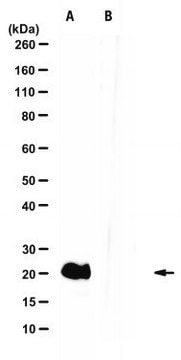
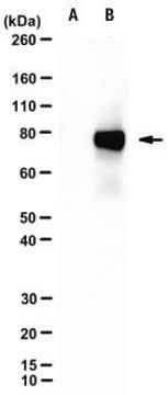
- Specificity: Clone MY-1 is a rat monoclonal antibody that detects Tyrosine-protein phosphatase non-receptor type substrate 1 (SIRPA/CD172a).
- Immunogen: Eosinophil-enriched cell fraction from the mouse small intestinal lamina propria.
- Application: Quality Control TestingEvaluated by Western Blotting in Mouse brain tissue extracts.Western Blotting Analysis: A 1:500 dilution of this antibody detected SIRPA in Mouse brain tissue extracts.Tested applicationsFlow Cytometry Analysis: A representative lot detected SIRPA in Flow Cytometry applications (Verjan Garcia, N., et al. (2011). J Immunol. 187(5):2268-77; Yanagita, T., et al. (2017). JCI Insight. 2(1); e89140). Immunofluorescence Analysis: A representative lot detected SIRPA in Immunofluorescence applications (Verjan Garcia, N., et al. (2011). J Immunol.;187(5):2268-77; Yanagita, T., et al. (2017). JCI Insight. 2(1):e89140).Inhibition Assay: A representative lot inhibited the growth of subcutaneously transplanted cancer cells in murine models. (Yanagita, T., et al. (2017). JCI Insight.;2(1):e89140; Sakamoto, M., et al. (2022). Proc Natl Acad Sci USA.;119(1)e2109923118).Immunoprecipitation Analysis: A representative lot immunoprecipitated SIRPA in Immunoprecipitation applications (Verjan Garcia, N., et al. (2011). J Immunol. 187(5):2268-77; Yanagita, T., et al. (2017). JCI Insight. 2(1):e89140).Western Blotting Analysis: A representative lot detected SIRPA in Western Blotting applications (Verjan Garcia, N., et al. (2011). J Immunol. 187(5):2268-77; Yanagita, T., et al. (2017). JCI Insight. 2(1):e89140).Note: Actual optimal working dilutions must be determined by end user as specimens, and experimental conditions may vary with the end user
- Physical form: Purified rat monoclonal antibody IgG2a in PBS without preservatives.
- Storage and Stability: Store at -10°C to -25°C. Handling Recommendations: Upon receipt and prior to removing the cap, centrifuge the vial and gently mix the solution. Aliquot into microcentrifuge tubes and store at -20°C. Avoid repeated freeze/thaw cycles, which may damage IgG and affect product performance.
- Other Notes: Concentration: Please refer to the Certificate of Analysis for the lot-specific concentration.
- Disclaimer: Unless otherwise stated in our catalog or other company documentation accompanying the product(s), our products are intended for research use only and are not to be used for any other purpose, which includes but is not limited to, unauthorized commercial uses, in vitro diagnostic uses, ex vivo or in vivo therapeutic uses or any type of consumption or application to humans or animals.
SAFETY INFORMATION
WGK
WGK 2
Flash Point(F)
Not applicable
Flash Point(C)
Not applicable
Compare Similar Items
Show Difference
biological source: rat
Quality Level: 200
conjugate: unconjugated
antibody form: purified antibody
antibody product type: primary antibodies
clone: MY-1, monoclonal
mol wt: calculated mol wt 56.14 kDaobserved mol wt ~95 kDa
purified by: using protein G
species reactivity: mouse
packaging: antibody small pack of 100 μL
biological source:
rat
Quality Level:
200
conjugate:
unconjugated
antibody form:
purified antibody
antibody product type:
primary antibodies
clone:
MY-1, monoclonal
mol wt:
calculated mol wt 56.14 kDaobserved mol wt ~95 kDa
purified by:
using protein G
species reactivity:
mouse
packaging:
antibody small pack of 100 μL
biological source: mouse
Quality Level: 200
conjugate: __
antibody form: purified antibody
antibody product type: primary antibodies
clone: GARP-50, monoclonal
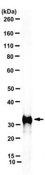
mol wt: calculated mol wt 31.73 kDaobserved mol wt ~32 kDa
purified by: using protein G
species reactivity: human
packaging: antibody small pack of 100 μL
biological source:
mouse
Quality Level:
200
conjugate:
__
antibody form:
purified antibody
antibody product type:
primary antibodies
clone:
GARP-50, monoclonal
mol wt:
calculated mol wt 31.73 kDaobserved mol wt ~32 kDa
purified by:
using protein G
species reactivity:
human
packaging:
antibody small pack of 100 μL

biological source: __
Quality Level: __
conjugate: __
antibody form: __
antibody product type: __
clone: __
mol wt: __
purified by: __
species reactivity: __
packaging: __
biological source:
__
Quality Level:
__
conjugate:
__
antibody form:
__
antibody product type:
__
clone:
__
mol wt:
__
purified by:
__
species reactivity:
__
packaging:
__