Rb Monoclonal Antibody (1F8), MonoMAb™, Zeta
Manufacturer: ZETA CORPORATION
Select a Size
| Pack Size | SKU | Availability | Price |
|---|---|---|---|
| Each of 1 | 50-223-8174-Each-of-1 | In Stock | ₹ 7,387.00 |
50-223-8174 - Each of 1
In Stock
Quantity
1
Base Price: ₹ 7,387.00
GST (18%): ₹ 1,329.66
Total Price: ₹ 8,716.66
Antigen
Rb
Classification
Monoclonal
Concentration
200 μg/mL
Formulation
tris with BSA, NP-40 and <0.1% sodium azide
Gene Accession No.
P06400
Gene Symbols
RB1
Immunogen
Human Rb1 protein fragment of 283 amino acid residues
Quantity
100 μL
Primary or Secondary
Primary
Target Species
Human
Product Type
Antibody
Isotype
IgG1 κ
Applications
Immunohistochemistry (Paraffin)
Clone
1F8
Conjugate
Unconjugated
Gene
RB1
Gene Alias
Cleaved Rb; delta RB-p70; DRb-p70; exon 17 tumor GOS561 substitution mutation causes premature stop; GOS563 exon 17 substitution mutation causes premature stop; OSRC; p104; p104 chicken Rb; P105RB; p105-Rb; Phospho-Retinoblastoma; pp105; pp110; PPP1R130; pRb; prepro-retinoblastoma-associated protein; protein phosphatase 1, regulatory subunit 130; rb; RB transcriptional corepressor 1; RB1; Rb-1; Rb-p70; Retin; Retinoblastoma; retinoblastoma 1; retinoblastoma 1 (including osteosarcoma); retinoblastoma 1 protein; retinoblastoma gene; retinoblastoma isoform A; retinoblastoma protein; retinoblastoma suspectibility protein; retinoblastoma tumor suppressor; retinoblastoma-associated protein
Host Species
Mouse
Purification Method
Protein A
Regulatory Status
RUO
Gene ID (Entrez)
5925
Content And Storage
2-8°C
Form
Liquid
Related Products


Description
- This product is diluted and in a ready-to-use formulation
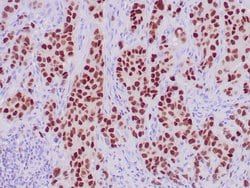
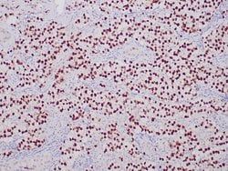
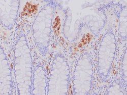
- A recommended positive control tissue for this product is Colon cancer, however positive controls are not limited to this tissue type
- The primary antibody is intended for laboratory professional use in the detection of the corresponding protein in formalin-fixed, paraffin-embedded tissue stained in manual qualitative immunohistochemistry (IHC) testing
- This antibody is intended to be used after the primary diagnosis of tumor has been made by conventional histopathology using non-immunological histochemical stains
- Antibody is used with formalin-fixed and paraffin-embedded sections
- Pretreatment of deparaffinized tissue with heat-induced epitope retrieval or enzymatic retrieval is recommended
- In general, immunohistochemical (IHC) staining techniques allow for the visualization of antigens via the sequential application of a specific antibody to the antigen (primary antibody), a secondary antibody to the primary antibody (link antibody), an enzyme complex and a chromogenic substrate with interposed washing steps
- The enzymatic activation of the chromogen results in a visible reaction product at the antigen site
- Results are interpreted using a light microscope and aid in the differential diagnosis of pathophysiological processes, which may or may not be associated with a particular antigen
- A positive tissue control must be run with every staining procedure performed
- This tissue may contain both positive and negative staining cells or tissue components and serve as both the positive and negative control tissue
- External Positive control materials should be fresh autopsy/biopsy/surgical specimens fixed, processed and embedded as soon as possible in the same manner as the patient sample (s)
- Positive tissue controls are indicative of correctly prepared tissues and proper staining methods
- The tissues used for the external positive control materials should be selected from the patient specimens with well-characterized low levels of the positive target activity that gives weak positive staining
- The low level of positivity for external positive controls is designed to ensure detection of subtle changes in the primary antibody sensitivity from instability or problems with the staining methodology
- A tissue with weak positive staining is more suitable for optimal quality control and for detecting minor levels of reagent degradation
- Internal or external negative control tissue may be used depending on the guidelines and policies that govern the organization to which the end user belongs to
- The variety of cell types present in many tissue sections offers internal negative control sites, but this should be verified by the user
- The components that do not stain should demonstrate the absence of specific staining, and provide an indication of non-specific background staining
- If specific staining occurs in the negative tissue control sites, results with the patient specimens must be considered invalid.
Compare Similar Items
Show Difference

Antigen: Rb
Classification: Monoclonal
Concentration: 200 μg/mL
Formulation: tris with BSA, NP-40 and <0.1% sodium azide
Gene Accession No.: P06400
Gene Symbols: RB1
Immunogen: Human Rb1 protein fragment of 283 amino acid residues
Quantity: 100 μL
Primary or Secondary: Primary
Target Species: Human
Product Type: Antibody
Isotype: IgG1 κ
Applications: Immunohistochemistry (Paraffin)
Clone: 1F8
Conjugate: Unconjugated
Gene: RB1
Gene Alias: Cleaved Rb; delta RB-p70; DRb-p70; exon 17 tumor GOS561 substitution mutation causes premature stop; GOS563 exon 17 substitution mutation causes premature stop; OSRC; p104; p104 chicken Rb; P105RB; p105-Rb; Phospho-Retinoblastoma; pp105; pp110; PPP1R130; pRb; prepro-retinoblastoma-associated protein; protein phosphatase 1, regulatory subunit 130; rb; RB transcriptional corepressor 1; RB1; Rb-1; Rb-p70; Retin; Retinoblastoma; retinoblastoma 1; retinoblastoma 1 (including osteosarcoma); retinoblastoma 1 protein; retinoblastoma gene; retinoblastoma isoform A; retinoblastoma protein; retinoblastoma suspectibility protein; retinoblastoma tumor suppressor; retinoblastoma-associated protein
Host Species: Mouse
Purification Method: Protein A
Regulatory Status: RUO
Gene ID (Entrez): 5925
Content And Storage: 2-8°C
Form: Liquid
Antigen: SALL4
Classification: Recombinant Monoclonal
Concentration: 100 μg/mL
Formulation: tris with BSA, NP-40 and <0.1% sodium azide
Gene Accession No.: Q9UJQ4
Gene Symbols: SALL4
Immunogen: Recombinant fragment (around aa1-185) of human MIC2 protein
Quantity: 100 μL
Primary or Secondary: Primary
Target Species: Human
Product Type: Antibody
Isotype: IgG
Applications: Immunohistochemistry (Paraffin)
Clone: ZR276
Conjugate: Unconjugated
Gene: SALL4
Gene Alias: 5730441M18Rik; AA407717; AL022809; AW536104; C330011P20Rik; C78083; C78563; dJ1112F19.1; DRRS; HSAL4; SALL4; sal-like 4; sal-like 4 (Drosophila); sal-like protein 4; spalt like transcription factor 4; spalt-like transcription factor 4; Tex20; Zinc finger protein 797; zinc finger protein SALL4; ZNF797
Host Species: Rabbit
Purification Method: Protein A
Regulatory Status: RUO
Gene ID (Entrez): 57167
Content And Storage: 2-8°C
Form: Liquid
Antigen: CD68
Classification: Recombinant Monoclonal
Concentration: 1 μg/mL
Formulation: tris with BSA, NP-40 and <0.1% sodium azide
Gene Accession No.: P34810
Gene Symbols: Cd68
Immunogen: Recombinant fragment (around aa1-200) of human CD68
Quantity: 7 mL
Primary or Secondary: Primary
Target Species: Human
Product Type: Antibody
Isotype: IgG
Applications: Immunohistochemistry (Paraffin)
Clone: ZR302
Conjugate: Unconjugated
Gene: Cd68
Gene Alias: CD68; CD68 antigen; CD68 molecule; GP110; Lamp4; macrophage antigen CD68; macrosialin; Scard1; scavenger receptor class D, member 1; similar to CD68 antigen
Host Species: Rabbit
Purification Method: Protein A
Regulatory Status: RUO
Gene ID (Entrez): 968
Content And Storage: 2-8°C
Form: Liquid