Cyclin B1 Antibody (SPM619), Novus Biologicals™
Mouse Monoclonal Antibody
Manufacturer: Fischer Scientific
The price for this product is unavailable. Please request a quote
Antigen
Cyclin B1
Concentration
0.2mg/mL
Applications
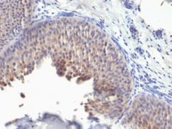
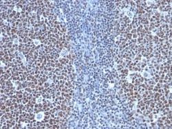
Flow Cytometry, Immunohistochemistry (Paraffin), Immunofluorescence
Conjugate
Unconjugated
Host Species
Mouse
Research Discipline
Cancer, Cell Biology, Cell Cycle and Replication, Mitotic Regulators, Tumor Suppressors
Formulation
10mM PBS and 0.05% BSA with 0.05% Sodium Azide
Gene ID (Entrez)
891
Immunogen
Recombinant human full-length CCNB1 protein
Primary or Secondary
Primary
Content And Storage
Store at 4C.
Clone
SPM619
Dilution
Flow Cytometry 0.5 - 1 ug/million cells in 0.1 ml, Immunohistochemistry-Paraffin 0.5 - 1.0 ug/ml, Immunofluorescence 1 - 2 ug/ml
Classification
Monoclonal
Form
Purified
Regulatory Status
RUO
Target Species
Human, Mouse
Gene Alias
CCNB, cyclin B1, G2/mitotic-specific cyclin B1, G2/mitotic-specific cyclin-B1
Gene Symbols
CCNB1
Isotype
IgG1 κ
Purification Method
Protein A or G purified
Test Specificity
It recognizes a protein of 55-62kDa, identified as cyclin B1. In mammals, cyclin B associates with inactive p34cdc2, which facilitates phosphorylation of p34cdc2 at aa 14Thr and 15Tyr. This maintains the inactive state until the end of G2-phase. The inactive cyclin B-p34cdc2 complex continues to accumulate in the cytoplasm until the completion of DNA synthesis, when Cdc25, a specific protein phosphatase, dephosphorylates aa 14Thr and 15Tyr of p34cdc2 rendering the complex active at the G2/M boundary. This mitotic kinase complex remains active until the metaphase/anaphase transition when cyclin B is degraded. This degradation process is ubiquitin-dependent and is necessary for the cell to exit mitosis. So, cyclin B-p34cdc2 plays a critical role in G2 to M transition.
Related Products


Description
- Ensure accurate, reproducible results in Flow Cytometry, Immunohistochemistry (Paraffin), Immunofluorescence Cyclin B1 Monoclonal specifically detects Cyclin B1 in Human, Mouse samples
- It is validated for Flow Cytometry, Immunohistochemistry, Immunocytochemistry/Immunofluorescence, Immunohistochemistry-Paraffin, Immunofluorescence.
Compare Similar Items
Show Difference
Antigen: Cyclin B1
Concentration: 0.2mg/mL
Applications: Flow Cytometry, Immunohistochemistry (Paraffin), Immunofluorescence
Conjugate: Unconjugated
Host Species: Mouse
Research Discipline: Cancer, Cell Biology, Cell Cycle and Replication, Mitotic Regulators, Tumor Suppressors
Formulation: 10mM PBS and 0.05% BSA with 0.05% Sodium Azide
Gene ID (Entrez): 891
Immunogen: Recombinant human full-length CCNB1 protein
Primary or Secondary: Primary
Content And Storage: Store at 4C.
Clone: SPM619
Dilution: Flow Cytometry 0.5 - 1 ug/million cells in 0.1 ml, Immunohistochemistry-Paraffin 0.5 - 1.0 ug/ml, Immunofluorescence 1 - 2 ug/ml
Classification: Monoclonal
Form: Purified
Regulatory Status: RUO
Target Species: Human, Mouse
Gene Alias: CCNB, cyclin B1, G2/mitotic-specific cyclin B1, G2/mitotic-specific cyclin-B1
Gene Symbols: CCNB1
Isotype: IgG1 κ
Purification Method: Protein A or G purified
Test Specificity: It recognizes a protein of 55-62kDa, identified as cyclin B1. In mammals, cyclin B associates with inactive p34cdc2, which facilitates phosphorylation of p34cdc2 at aa 14Thr and 15Tyr. This maintains the inactive state until the end of G2-phase. The inactive cyclin B-p34cdc2 complex continues to accumulate in the cytoplasm until the completion of DNA synthesis, when Cdc25, a specific protein phosphatase, dephosphorylates aa 14Thr and 15Tyr of p34cdc2 rendering the complex active at the G2/M boundary. This mitotic kinase complex remains active until the metaphase/anaphase transition when cyclin B is degraded. This degradation process is ubiquitin-dependent and is necessary for the cell to exit mitosis. So, cyclin B-p34cdc2 plays a critical role in G2 to M transition.
Antigen: Cytochrome c
Concentration: 0.2mg/mL
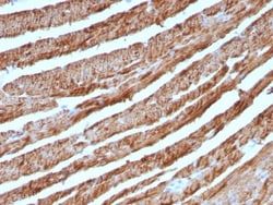
Applications: Western Blot, Flow Cytometry, Immunohistochemistry (Paraffin), Immunofluorescence
Conjugate: Unconjugated
Host Species: Mouse
Research Discipline: Apoptosis, Cellular Markers, Cholesterol Metabolism, Core ESC Like Genes, Lipid and Metabolism, Mitochondrial Markers, Stem Cell Markers
Formulation: 1.0mM PBS and 0.05% BSA with 0.05% Sodium Azide
Gene ID (Entrez): 54205
Immunogen: Recombinant cytochrome c protein
Primary or Secondary: Primary
Content And Storage: Store at 4C.
Clone: CTC05
Dilution: Western Blot 0.5 - 1.0 ug/ml, Flow Cytometry 0.5 - 1 ug/million cells in 0.1 ml, Immunohistochemistry-Paraffin 0.25 - 0.5 ug/ml, Immunofluorescence 0.5 - 1.0 ug/ml
Classification: Monoclonal
Form: Purified
Regulatory Status: RUO
Target Species: Human, Mouse, Rat, Amphibian, Avian, Canine, Drosophila, Equine
Gene Alias: CYCHCS, cytochrome c, cytochrome c, somatic, THC4
Gene Symbols: CYCS
Isotype: IgG2b κ
Purification Method: Protein A or G purified
Test Specificity: Cytochrome C is a well-characterized mobile electron transport protein that is essential to energy conversion in all aerobic organisms. In mammalian cells, this highly conserved protein is normally localized to the mitochondrial inter-membrane space. More recent studies have identified cytosolic cytochrome c as a factor necessary for activation of apoptosis. During apoptosis, cytochrome c is trans-located from the mitochondrial membrane to the cytosol, where it is required for activation of caspase-3 (CPP32). Overexpression of Bcl-2 has been shown to prevent the translocation of cytochrome c, thereby blocking the apoptotic process. Overexpression of Bax has been shown to induce the release of cytochrome c and to induce cell death. The release of cytochrome c from the mitochondria is thought to trigger an apoptotic cascade, whereby Apaf-1 binds to Apaf-3 (caspase-9) in a cytochrome c-dependent manner, leading to caspase-9 cleavage of caspase-3.
Antigen: Cytochrome c
Concentration: 0.2mg/mL
Applications: Western Blot, Flow Cytometry, Immunohistochemistry (Paraffin), Immunofluorescence
Conjugate: Unconjugated
Host Species: Mouse
Research Discipline: Apoptosis, Cellular Markers, Cholesterol Metabolism, Core ESC Like Genes, Lipid and Metabolism, Mitochondrial Markers, Stem Cell Markers
Formulation: 1.0mM PBS and 0.05% BSA with 0.05% Sodium Azide
Gene ID (Entrez): 54205
Immunogen: Recombinant cytochrome c protein
Primary or Secondary: Primary
Content And Storage: Store at 4C.
Clone: CTC05
Dilution: Western Blot 0.5 - 1.0 ug/ml, Flow Cytometry 0.5 - 1 ug/million cells in 0.1 ml, Immunohistochemistry-Paraffin 0.25 - 0.5 ug/ml, Immunofluorescence 0.5 - 1.0 ug/ml
Classification: Monoclonal
Form: Purified
Regulatory Status: RUO
Target Species: Human, Mouse, Rat, Amphibian, Avian, Canine, Drosophila, Equine
Gene Alias: CYCHCS, cytochrome c, cytochrome c, somatic, THC4
Gene Symbols: CYCS
Isotype: IgG2b κ
Purification Method: Protein A or G purified
Test Specificity: Cytochrome C is a well-characterized mobile electron transport protein that is essential to energy conversion in all aerobic organisms. In mammalian cells, this highly conserved protein is normally localized to the mitochondrial inter-membrane space. More recent studies have identified cytosolic cytochrome c as a factor necessary for activation of apoptosis. During apoptosis, cytochrome c is trans-located from the mitochondrial membrane to the cytosol, where it is required for activation of caspase-3 (CPP32). Overexpression of Bcl-2 has been shown to prevent the translocation of cytochrome c, thereby blocking the apoptotic process. Overexpression of Bax has been shown to induce the release of cytochrome c and to induce cell death. The release of cytochrome c from the mitochondria is thought to trigger an apoptotic cascade, whereby Apaf-1 binds to Apaf-3 (caspase-9) in a cytochrome c-dependent manner, leading to caspase-9 cleavage of caspase-3.