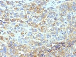
DPPIV/CD26 Antibody (134-2C2), Novus Biologicals™
Mouse Monoclonal Antibody
Manufacturer: Fischer Scientific
The price for this product is unavailable. Please request a quote
Antigen
DPPIV/CD26
Concentration
0.2 mg/mL
Applications
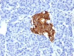
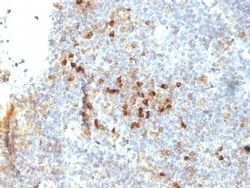
Flow Cytometry, Immunohistochemistry (Frozen), Immunofluorescence
Conjugate
Unconjugated
Host Species
Mouse
Research Discipline
Adaptive Immunity, Cellular Markers, GPCR, Immunology
Formulation
10mM PBS and 0.05% BSA with 0.05% Sodium Azide
Gene Alias
ADABP, ADCP-2, ADCP2DPP IV, Adenosine deaminase complexing protein 2TP103, CD26 antigen, CD26T-cell activation antigen CD26, dipeptidyl peptidase 4, Dipeptidyl peptidase IV, dipeptidylpeptidase 4, dipeptidyl-peptidase 4, dipeptidylpeptidase IV (CD26, adenosine deaminase complexing protein 2), DPPIV, EC 3.4.14.5
Gene Symbols
DPP4
Isotype
IgM κ
Purification Method
Protein A or G purified
Test Specificity
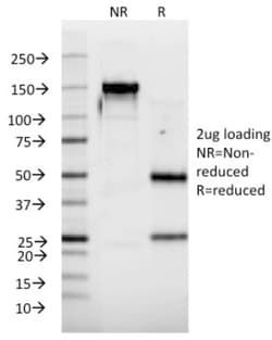
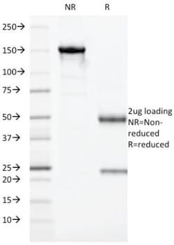
Recognizes a glycoprotein of 110kDa, identified as CD26. It is an atypical serine protease belonging to the prolyl oligopeptidase family. It is expressed on lymphocyte cells and is upregulated during T-cell activation. CD26 is also expressed on activated B cells and natural killer cells and abundantly on epithelia. CD26 is implicated in a variety of biological functions including T-cell activation, cell adhesion with extracellular matrix such as fibronectin or collagens, and in HIV infection.
Clone
134-2C2
Dilution
Flow Cytometry 0.5 - 1 ug/million cells in 0.1 ml, Immunohistochemistry-Frozen 0.5 - 1.0 ug/ml, Immunofluorescence 0.5 - 1.0 ug/ml
Classification
Monoclonal
Form
Purified
Regulatory Status
RUO
Target Species
Human
Gene Accession No.
P27487
Gene ID (Entrez)
1803
Immunogen
A synthetic peptide from the amino terminal region of CD26.
Primary or Secondary
Primary
Content And Storage
Store at 4C.
Molecular Weight of Antigen
110 kDa
Related Products


Description
- Ensure accurate, reproducible results in Flow Cytometry, Immunohistochemistry (Frozen), Immunofluorescence DPPIV/CD26 Monoclonal specifically detects DPPIV/CD26 in Human samples
- It is validated for Flow Cytometry, Immunohistochemistry, Immunocytochemistry/Immunofluorescence, Immunohistochemistry-Frozen, Immunofluorescence.
Compare Similar Items
Show Difference
Antigen: DPPIV/CD26
Concentration: 0.2 mg/mL
Applications: Flow Cytometry, Immunohistochemistry (Frozen), Immunofluorescence
Conjugate: Unconjugated
Host Species: Mouse
Research Discipline: Adaptive Immunity, Cellular Markers, GPCR, Immunology
Formulation: 10mM PBS and 0.05% BSA with 0.05% Sodium Azide
Gene Alias: ADABP, ADCP-2, ADCP2DPP IV, Adenosine deaminase complexing protein 2TP103, CD26 antigen, CD26T-cell activation antigen CD26, dipeptidyl peptidase 4, Dipeptidyl peptidase IV, dipeptidylpeptidase 4, dipeptidyl-peptidase 4, dipeptidylpeptidase IV (CD26, adenosine deaminase complexing protein 2), DPPIV, EC 3.4.14.5
Gene Symbols: DPP4
Isotype: IgM κ
Purification Method: Protein A or G purified
Test Specificity: Recognizes a glycoprotein of 110kDa, identified as CD26. It is an atypical serine protease belonging to the prolyl oligopeptidase family. It is expressed on lymphocyte cells and is upregulated during T-cell activation. CD26 is also expressed on activated B cells and natural killer cells and abundantly on epithelia. CD26 is implicated in a variety of biological functions including T-cell activation, cell adhesion with extracellular matrix such as fibronectin or collagens, and in HIV infection.
Clone: 134-2C2
Dilution: Flow Cytometry 0.5 - 1 ug/million cells in 0.1 ml, Immunohistochemistry-Frozen 0.5 - 1.0 ug/ml, Immunofluorescence 0.5 - 1.0 ug/ml
Classification: Monoclonal
Form: Purified
Regulatory Status: RUO
Target Species: Human
Gene Accession No.: P27487
Gene ID (Entrez): 1803
Immunogen: A synthetic peptide from the amino terminal region of CD26.
Primary or Secondary: Primary
Content And Storage: Store at 4C.
Molecular Weight of Antigen: 110 kDa
Antigen: DPPIV/CD26
Concentration: 0.2 mg/mL
Applications: Flow Cytometry, Immunohistochemistry (Frozen), Immunofluorescence
Conjugate: Unconjugated
Host Species: Mouse
Research Discipline: Adaptive Immunity, Cellular Markers, GPCR, Immunology
Formulation: 10mM PBS and 0.05% BSA with 0.05% Sodium Azide
Gene Alias: ADABP, ADCP-2, ADCP2DPP IV, Adenosine deaminase complexing protein 2TP103, CD26 antigen, CD26T-cell activation antigen CD26, dipeptidyl peptidase 4, Dipeptidyl peptidase IV, dipeptidylpeptidase 4, dipeptidyl-peptidase 4, dipeptidylpeptidase IV (CD26, adenosine deaminase complexing protein 2), DPPIV, EC 3.4.14.5
Gene Symbols: DPP4
Isotype: IgG2b κ
Purification Method: Protein A or G purified
Test Specificity: Recognizes a glycoprotein of 110kDa, identified as CD26 (Workshop VI; Code: N-L039). It is an atypical serine protease belonging to the prolyl oligopeptidase family. It is expressed on lymphocyte cells and is upregulated during T-cell activation. CD26 is also expressed on activated B cells and natural killer cells and abundantly on epithelia. CD26 is implicated in a variety of biological functions including T-cell activation, cell adhesion with extracellular matrix such as fibronectin or collagens, and in HIV infection. Cross-linking of CD26 using this antibody dramatically enhances the anti-CD3-induced IL-2 production. In Western blotting, this MAb reacts with only glycosylated CD26, but not with the deglycosylated form. It does not prevent ADA binding to CD26.
Clone: 202.36
Dilution: Flow Cytometry 0.5 - 1 ug/million cells in 0.1 ml, Immunohistochemistry-Frozen 0.5 - 1.0 ug/ml, Immunofluorescence 0.5 - 1.0 ug/ml
Classification: Monoclonal
Form: Purified
Regulatory Status: RUO
Target Species: Human, Rat, Porcine (Negative), Sheep (Negative)
Gene Accession No.: P27487
Gene ID (Entrez): 1803
Immunogen: Human T cell clone
Primary or Secondary: Primary
Content And Storage: Store at 4C.
Molecular Weight of Antigen: 110 kDa
Antigen: ICAM-1/CD54
Concentration: 0.2mg/mL
Applications: Flow Cytometry, Immunohistochemistry (Paraffin), SDS-Page, Immunofluorescence
Conjugate: Unconjugated
Host Species: Mouse
Research Discipline: Adaptive Immunity, Cancer, Cell Biology, Immunology, Mesenchymal Stem Cell Markers, Stem Cell Markers
Formulation: 10mM PBS and 0.05% BSA with 0.05% Sodium Azide
Gene Alias: BB2, CD54, CD54 antigen, cell surface glycoprotein P3.58, human rhinovirus receptor, ICAM-1, intercellular adhesion molecule 1, intercellular adhesion molecule 1 (CD54), human rhinovirus receptor, Major group rhinovirus receptor, P3.58
Gene Symbols: ICAM1
Isotype: IgG2b κ
Purification Method: Protein A or G purified
Test Specificity: Recognizes an 85-115kDa protein (variation with cell type), identified as intercellular adhesion molecule (ICAM-1) (Workshop IV). It has 7 potential N-linked glycosylation sites. ICAM-1 is a single chain glycoprotein of Ig supergene family, present on unstimulated endothelial cells (EC) and on a variety of other cell types including activated fibroblasts, EC, macrophages, and lymphocytes. ICAM-1 mediates cell adhesion by binding to integrins CD11a/CD18 (leukocyte adhesion molecule, LFA-1) and to CD11b/CD18 (Mac-1). This interaction enhances antigen-specific T-cell activation. ICAM-1 also binds to CD43 and to Plasmodium falciparum infected RBCs. W-CAM-1 MAb blocks aggregation of cell lines mediated by the ICAM-1 and blocks homotypic binding of purified populations of activated T- and B-lymphocytes and also aggregation of mixed T- and B-cell blasts. It inhibits T-cell adhesion to normal human endothelial cells. Activation induced by cell-cell contact (mixed lymphocyte reaction, T-cell me
Clone: W-CAM-1 (same as Wehi-CAM-1 or 1H4)
Dilution: Flow Cytometry 0.5 - 1 ug/million cells in 0.1 ml, Immunohistochemistry-Paraffin 2 - 4 ug/ml, SDS-Page, Immunofluorescence 0.5 - 1.0 ug/ml
Classification: Monoclonal
Form: Purified
Regulatory Status: RUO
Target Species: Human
Gene Accession No.: __
Gene ID (Entrez): 3383
Immunogen: Raji Burkitt lymphoma cells
Primary or Secondary: Primary
Content And Storage: Store at 4C.
Molecular Weight of Antigen: __