DPPIV/CD26 Antibody (202.36), Novus Biologicals™
Mouse Monoclonal Antibody
Manufacturer: Fischer Scientific
The price for this product is unavailable. Please request a quote
Antigen
DPPIV/CD26
Concentration
0.2 mg/mL
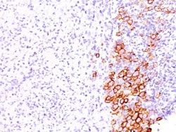
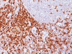
Applications
Flow Cytometry, Immunohistochemistry (Frozen), Immunofluorescence
Conjugate
Unconjugated
Host Species
Mouse
Research Discipline
Adaptive Immunity, Cellular Markers, GPCR, Immunology
Formulation
10mM PBS and 0.05% BSA with 0.05% Sodium Azide
Gene Alias
ADABP, ADCP-2, ADCP2DPP IV, Adenosine deaminase complexing protein 2TP103, CD26 antigen, CD26T-cell activation antigen CD26, dipeptidyl peptidase 4, Dipeptidyl peptidase IV, dipeptidylpeptidase 4, dipeptidyl-peptidase 4, dipeptidylpeptidase IV (CD26, adenosine deaminase complexing protein 2), DPPIV, EC 3.4.14.5
Gene Symbols
DPP4
Isotype
IgG2b κ
Purification Method
Protein A or G purified
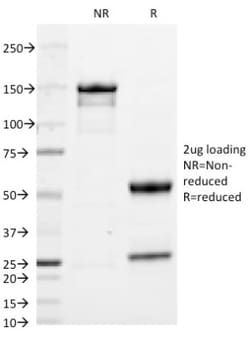
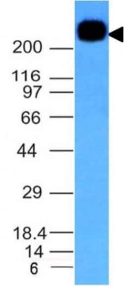
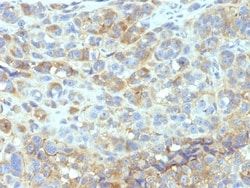
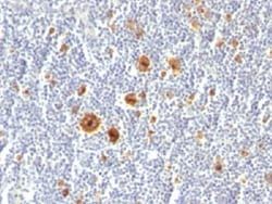
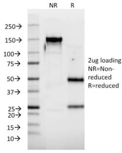
Test Specificity
Recognizes a glycoprotein of 110kDa, identified as CD26 (Workshop VI; Code: N-L039). It is an atypical serine protease belonging to the prolyl oligopeptidase family. It is expressed on lymphocyte cells and is upregulated during T-cell activation. CD26 is also expressed on activated B cells and natural killer cells and abundantly on epithelia. CD26 is implicated in a variety of biological functions including T-cell activation, cell adhesion with extracellular matrix such as fibronectin or collagens, and in HIV infection. Cross-linking of CD26 using this antibody dramatically enhances the anti-CD3-induced IL-2 production. In Western blotting, this MAb reacts with only glycosylated CD26, but not with the deglycosylated form. It does not prevent ADA binding to CD26.
Clone
202.36
Dilution
Flow Cytometry 0.5 - 1 ug/million cells in 0.1 ml, Immunohistochemistry-Frozen 0.5 - 1.0 ug/ml, Immunofluorescence 0.5 - 1.0 ug/ml
Classification
Monoclonal
Form
Purified
Regulatory Status
RUO
Target Species
Human, Rat, Porcine (Negative), Sheep (Negative)
Gene Accession No.
P27487
Gene ID (Entrez)
1803
Immunogen
Human T cell clone
Primary or Secondary
Primary
Content And Storage
Store at 4C.
Molecular Weight of Antigen
110 kDa
Related Products

Description
- Ensure accurate, reproducible results in Flow Cytometry, Immunohistochemistry (Frozen), Immunofluorescence DPPIV/CD26 Monoclonal specifically detects DPPIV/CD26 in Human, Rat, Porcine (Negative), Sheep (Negative) samples
- It is validated for Flow Cytometry, Immunohistochemistry, Immunocytochemistry/Immunofluorescence, Immunohistochemistry-Frozen, Immunofluorescence.
Compare Similar Items
Show Difference
Antigen: DPPIV/CD26
Concentration: 0.2 mg/mL
Applications: Flow Cytometry, Immunohistochemistry (Frozen), Immunofluorescence
Conjugate: Unconjugated
Host Species: Mouse
Research Discipline: Adaptive Immunity, Cellular Markers, GPCR, Immunology
Formulation: 10mM PBS and 0.05% BSA with 0.05% Sodium Azide
Gene Alias: ADABP, ADCP-2, ADCP2DPP IV, Adenosine deaminase complexing protein 2TP103, CD26 antigen, CD26T-cell activation antigen CD26, dipeptidyl peptidase 4, Dipeptidyl peptidase IV, dipeptidylpeptidase 4, dipeptidyl-peptidase 4, dipeptidylpeptidase IV (CD26, adenosine deaminase complexing protein 2), DPPIV, EC 3.4.14.5
Gene Symbols: DPP4
Isotype: IgG2b κ
Purification Method: Protein A or G purified
Test Specificity: Recognizes a glycoprotein of 110kDa, identified as CD26 (Workshop VI; Code: N-L039). It is an atypical serine protease belonging to the prolyl oligopeptidase family. It is expressed on lymphocyte cells and is upregulated during T-cell activation. CD26 is also expressed on activated B cells and natural killer cells and abundantly on epithelia. CD26 is implicated in a variety of biological functions including T-cell activation, cell adhesion with extracellular matrix such as fibronectin or collagens, and in HIV infection. Cross-linking of CD26 using this antibody dramatically enhances the anti-CD3-induced IL-2 production. In Western blotting, this MAb reacts with only glycosylated CD26, but not with the deglycosylated form. It does not prevent ADA binding to CD26.
Clone: 202.36
Dilution: Flow Cytometry 0.5 - 1 ug/million cells in 0.1 ml, Immunohistochemistry-Frozen 0.5 - 1.0 ug/ml, Immunofluorescence 0.5 - 1.0 ug/ml
Classification: Monoclonal
Form: Purified
Regulatory Status: RUO
Target Species: Human, Rat, Porcine (Negative), Sheep (Negative)
Gene Accession No.: P27487
Gene ID (Entrez): 1803
Immunogen: Human T cell clone
Primary or Secondary: Primary
Content And Storage: Store at 4C.
Molecular Weight of Antigen: 110 kDa
Antigen: ICAM-1/CD54
Concentration: 0.2mg/mL
Applications: Flow Cytometry, Immunohistochemistry (Paraffin), SDS-Page, Immunofluorescence
Conjugate: Unconjugated
Host Species: Mouse
Research Discipline: Adaptive Immunity, Cancer, Cell Biology, Immunology, Mesenchymal Stem Cell Markers, Stem Cell Markers
Formulation: 10mM PBS and 0.05% BSA with 0.05% Sodium Azide
Gene Alias: BB2, CD54, CD54 antigen, cell surface glycoprotein P3.58, human rhinovirus receptor, ICAM-1, intercellular adhesion molecule 1, intercellular adhesion molecule 1 (CD54), human rhinovirus receptor, Major group rhinovirus receptor, P3.58
Gene Symbols: ICAM1
Isotype: IgG2b κ
Purification Method: Protein A or G purified
Test Specificity: Recognizes an 85-115kDa protein (variation with cell type), identified as intercellular adhesion molecule (ICAM-1) (Workshop IV). It has 7 potential N-linked glycosylation sites. ICAM-1 is a single chain glycoprotein of Ig supergene family, present on unstimulated endothelial cells (EC) and on a variety of other cell types including activated fibroblasts, EC, macrophages, and lymphocytes. ICAM-1 mediates cell adhesion by binding to integrins CD11a/CD18 (leukocyte adhesion molecule, LFA-1) and to CD11b/CD18 (Mac-1). This interaction enhances antigen-specific T-cell activation. ICAM-1 also binds to CD43 and to Plasmodium falciparum infected RBCs. W-CAM-1 MAb blocks aggregation of cell lines mediated by the ICAM-1 and blocks homotypic binding of purified populations of activated T- and B-lymphocytes and also aggregation of mixed T- and B-cell blasts. It inhibits T-cell adhesion to normal human endothelial cells. Activation induced by cell-cell contact (mixed lymphocyte reaction, T-cell me
Clone: W-CAM-1 (same as Wehi-CAM-1 or 1H4)
Dilution: Flow Cytometry 0.5 - 1 ug/million cells in 0.1 ml, Immunohistochemistry-Paraffin 2 - 4 ug/ml, SDS-Page, Immunofluorescence 0.5 - 1.0 ug/ml
Classification: Monoclonal
Form: Purified
Regulatory Status: RUO
Target Species: Human
Gene Accession No.: __
Gene ID (Entrez): 3383
Immunogen: Raji Burkitt lymphoma cells
Primary or Secondary: Primary
Content And Storage: Store at 4C.
Molecular Weight of Antigen: __
Antigen: IFN-alpha 2
Concentration: 0.2 mg/mL
Applications: Flow Cytometry, SDS-Page, Immunofluorescence
Conjugate: Unconjugated
Host Species: Mouse
Research Discipline: Apoptosis, Cytokine Research
Formulation: 10mM PBS and 0.05% BSA with 0.05% Sodium Azide
Gene Alias: alpha-2a interferon, IFNA, IFN-alpha-2, IFN-alphaA, INFA2, interferon alpha 2b, interferon alpha A, interferon alpha-2, Interferon alpha-A, interferon, alpha 2, LeIF A, MGC125764, MGC125765
Gene Symbols: IFNA2
Isotype: IgG1 κ
Purification Method: Protein A or G purified
Test Specificity: Recognizes a protein of 16-27kDa, identified as human interferon-II) (IFN-(II). Its epitope maps between aa112-148 of IFN-II) (total aa172). This MAb is specific for IFN-(II) and does not cross-react with IFN-(I). The site recognized by this MAb is called site I and is responsible for the antiviral and anti-proliferative activities of IFN-(II). Epitopes of N27 and N39 MAbs are different and represent a good combination of antibodies to set up an ELISA assay for the quantitation of IFN-(II) after viral infections. The IFN- family consists of 24 or more genes or pseudo-genes. IFN-II) is one of the two distinct families (I and II) of human IFN-. The -interferon are mainly produced by lymphocytes, monocytes, macrophages, and cell lines such as Namalwa and KG1 following induction by viruses, nucleic acids, and glucocorticoid hormones. They are involved in virus resistance on target cells, inhibition of cell proliferation, induction of cytokines and regulation of expression of MHC class I an
Clone: N39
Dilution: Flow Cytometry 0.5 - 1 ug/million cells in 0.1 ml, SDS-Page, Immunofluorescence 0.5 - 1.0 ug/ml
Classification: Monoclonal
Form: Purified
Regulatory Status: RUO
Target Species: Human
Gene Accession No.: __
Gene ID (Entrez): 3440
Immunogen: Purified recombinant human IFN-alpha2
Primary or Secondary: Primary
Content And Storage: Store at 4C.
Molecular Weight of Antigen: __