Involucrin Antibody (IVRN/827), Novus Biologicals™
Mouse Monoclonal Antibody
Manufacturer: Fischer Scientific
The price for this product is unavailable. Please request a quote
Antigen
Involucrin
Concentration
0.2mg/mL
Applications
Flow Cytometry, Immunohistochemistry (Paraffin), Immunofluorescence
Conjugate
Unconjugated
Host Species
Mouse
Research Discipline
Extracellular Matrix
Formulation
10mM PBS and 0.05% BSA with 0.05% Sodium Azide
Gene ID (Entrez)
3713
Immunogen
Purified involucrin from human keratinocytes
Primary or Secondary
Primary
Content And Storage
Store at 4C.
Clone
IVRN/827
Dilution
Flow Cytometry 0.5 - 1 ug/million cells in 0.1 ml, Immunohistochemistry-Paraffin 0.1 - 0.2 ug/ml, Immunofluorescence 1 - 2 ug/ml
Classification
Monoclonal
Form
Purified
Regulatory Status
RUO
Target Species
Human
Gene Alias
involucrin
Gene Symbols
IVL
Isotype
IgG1 κ
Purification Method
Protein A or G purified
Test Specificity
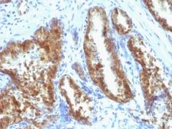
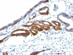
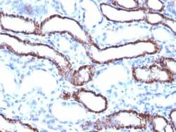
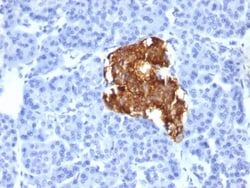
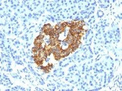
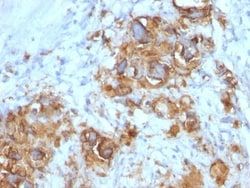
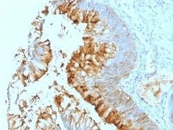
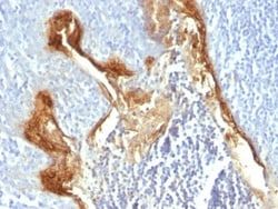
It recognizes a protein of 66kDa-170kDa, identified as involucrin. In Western blotting of cultured human keratinocytes, this MAb reacts with a 120kDa protein. Involucrin is expressed in a range of stratified squamous epithelia, including the cornea, which lacks a distinct cornified layer. In normal epidermis, it is first expressed in the upper spinous layers, and in keratinocyte cultures, all cells that have left the basal layer express it. Involucrin expression is altered in pathological conditions: in psoriasis and other benign epidermal hyperplasias, involucrin expression begins closer to the basal layer than normal; expression is abnormal in squamous cell carcinomas and premalignant lesions, and is reduced in severe dysplasias of the larynx and cervix.
Related Products
Description
- Ensure accurate, reproducible results in Flow Cytometry, Immunohistochemistry (Paraffin), Immunofluorescence Involucrin Monoclonal specifically detects Involucrin in Human samples
- It is validated for Flow Cytometry, Immunohistochemistry, Immunocytochemistry/Immunofluorescence, Immunohistochemistry-Paraffin, Immunofluorescence.
Compare Similar Items
Show Difference
Antigen: Involucrin
Concentration: 0.2mg/mL
Applications: Flow Cytometry, Immunohistochemistry (Paraffin), Immunofluorescence
Conjugate: Unconjugated
Host Species: Mouse
Research Discipline: Extracellular Matrix
Formulation: 10mM PBS and 0.05% BSA with 0.05% Sodium Azide
Gene ID (Entrez): 3713
Immunogen: Purified involucrin from human keratinocytes
Primary or Secondary: Primary
Content And Storage: Store at 4C.
Clone: IVRN/827
Dilution: Flow Cytometry 0.5 - 1 ug/million cells in 0.1 ml, Immunohistochemistry-Paraffin 0.1 - 0.2 ug/ml, Immunofluorescence 1 - 2 ug/ml
Classification: Monoclonal
Form: Purified
Regulatory Status: RUO
Target Species: Human
Gene Alias: involucrin
Gene Symbols: IVL
Isotype: IgG1 κ
Purification Method: Protein A or G purified
Test Specificity: It recognizes a protein of 66kDa-170kDa, identified as involucrin. In Western blotting of cultured human keratinocytes, this MAb reacts with a 120kDa protein. Involucrin is expressed in a range of stratified squamous epithelia, including the cornea, which lacks a distinct cornified layer. In normal epidermis, it is first expressed in the upper spinous layers, and in keratinocyte cultures, all cells that have left the basal layer express it. Involucrin expression is altered in pathological conditions: in psoriasis and other benign epidermal hyperplasias, involucrin expression begins closer to the basal layer than normal; expression is abnormal in squamous cell carcinomas and premalignant lesions, and is reduced in severe dysplasias of the larynx and cervix.

Antigen: Isocitrate Dehydrogenase 1/IDH1
Concentration: 0.2mg/mL
Applications: Western Blot, Flow Cytometry, Immunohistochemistry (Paraffin), Immunofluorescence
Conjugate: Unconjugated
Host Species: Mouse
Research Discipline: Stem Cell Markers
Formulation: 1.0mM PBS and 0.05% BSA with 0.05% Sodium Azide
Gene ID (Entrez): 3417
Immunogen: Recombinant fragment (119 Amino acid residues around aa 280-420) of human IDH1 protein
Primary or Secondary: Primary
Content And Storage: Store at 4C.
Clone: IDH1/1152
Dilution: Western Blot 0.5 - 1.0 ug/ml, Flow Cytometry 0.5 - 1 ug/million cells in 0.1 ml, Immunohistochemistry-Paraffin 0.5 - 1.0 ug/ml, Immunofluorescence 0.5 - 1.0 ug/ml
Classification: Monoclonal
Form: Purified
Regulatory Status: RUO
Target Species: Human
Gene Alias: Cytosolic NADP-isocitrate dehydrogenase, EC 1.1.1.42, IDCD, IDH, IDP, IDPC, isocitrate dehydrogenase [NADP] cytoplasmic, isocitrate dehydrogenase 1 (NADP+), soluble, NADP(+)-specific ICDH, NADP-dependent isocitrate dehydrogenase, cytosolic, NADP-dependent isocitrate dehydrogenase, peroxisomal, Oxalosuccinate decarboxylase, PICD
Gene Symbols: IDH1
Isotype: IgG1 κ
Purification Method: Protein A or G purified
Test Specificity: It recognizes a 45kDa protein, which is identified as isocitrate dehydrogenase (IDH1). It belongs to the isocitrate and isopropylmalate dehydrogenases family. IDH1 catalyzes the third step of the citric acid cycle, which involves the oxidative decarboxylation of isocitrate, forming ketoglutarate and CO2 in a two-step reaction. The first step involves the oxidation of isocitrate to the intermediate oxalosuccinate, while the second step involves the production of ketoglutarate. During this process, either NADH or NADPH is produced along with CO2. Recently, an inactivating mutation of IDH1 has been implicated in glioblastoma. IDH1 appears to function as a tumor suppressor that, when mutationally inactivated, contributes to tumorigenesis in part through induction of the HIF-1 pathway.