p53 Antibody (TRP/817), Novus Biologicals™
Mouse Monoclonal Antibody
Manufacturer: Fischer Scientific
The price for this product is unavailable. Please request a quote
Antigen
p53
Concentration
0.2mg/mL
Applications
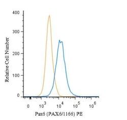
Western Blot, Flow Cytometry, Immunohistochemistry (Paraffin), Immunofluorescence, KnockDown
Conjugate
Unconjugated
Host Species
Mouse
Research Discipline
Apoptosis, Cancer, Cell Cycle and Replication, Cellular Markers, Checkpoint signaling, Core ESC Like Genes, DNA Double Strand Break Repair, DNA Repair, HIF Target Genes, Hypoxia, Neuroscience, Neurotransmission, p53 Pathway, Stem Cell Markers, Transcription Factors and Regulators, Tumor Suppressors
Formulation
10mM PBS and 0.05% BSA with 0.05% Sodium Azide
Gene ID (Entrez)
7157
Immunogen
Recombinant human TP53 protein
Primary or Secondary
Primary
Content And Storage
Store at 4C.
Molecular Weight of Antigen
53 kDa
Clone
TRP/817
Dilution
Western Blot 0.5 - 1.0 ug/ml, Flow Cytometry 0.5 - 1 ug/million cells in 0.1 ml, Immunohistochemistry-Paraffin 0.25 - 0.5 ug/ml, Immunofluorescence 0.5 - 1.0 ug/ml, Knockout Validated 0.5 ug/ml
Classification
Monoclonal
Form
Purified
Regulatory Status
RUO
Target Species
Human
Gene Alias
Antigen NY-CO-13, FLJ92943, LFS1TRP53, p53, p53 tumor suppressor, P53cellular tumor antigen p53, Phosphoprotein p53, transformation-related protein 53, tumor protein p53, Tumor suppressor p53
Gene Symbols
TP53
Isotype
IgG2b κ
Purification Method
Protein A or G purified
Test Specificity
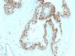
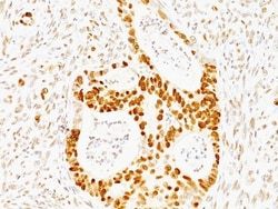
Recognizes a 53kDa protein, which is identified as p53 suppressor gene product. It reacts with the mutant as well as the wild form of p53 protein. p53 is a tumor suppressor gene expressed in a wide variety of tissue types and is involved in regulating cell growth, replication, and apoptosis. It binds to MDM2, SV40 T antigen and human papilloma virus E6 protein. Positive nuclear staining with p53 antibody has been reported to be a negative prognostic factor in breast carcinoma, lung carcinoma, colorectal, and urothelial carcinoma. Anti-p53 positivity has also been used to differentiate uterine serous carcinoma from endometrioid carcinoma as well as to detect intratubular germ cell neoplasia. Mutations involving p53 are found in a wide variety of malignant tumors, including breast, ovarian, bladder, colon, lung, and melanoma.
Related Products


Description
- Ensure accurate, reproducible results in Western Blot, Flow Cytometry, Immunohistochemistry (Paraffin), Immunofluorescence p53 Monoclonal specifically detects p53 in Human samples
- It is validated for Western Blot, Immunohistochemistry, Immunohistochemistry-Paraffin, Knockout Validated, Knockdown Validated.
Compare Similar Items
Show Difference
Antigen: p53
Concentration: 0.2mg/mL
Applications: Western Blot, Flow Cytometry, Immunohistochemistry (Paraffin), Immunofluorescence, KnockDown
Conjugate: Unconjugated
Host Species: Mouse
Research Discipline: Apoptosis, Cancer, Cell Cycle and Replication, Cellular Markers, Checkpoint signaling, Core ESC Like Genes, DNA Double Strand Break Repair, DNA Repair, HIF Target Genes, Hypoxia, Neuroscience, Neurotransmission, p53 Pathway, Stem Cell Markers, Transcription Factors and Regulators, Tumor Suppressors
Formulation: 10mM PBS and 0.05% BSA with 0.05% Sodium Azide
Gene ID (Entrez): 7157
Immunogen: Recombinant human TP53 protein
Primary or Secondary: Primary
Content And Storage: Store at 4C.
Molecular Weight of Antigen: 53 kDa
Clone: TRP/817
Dilution: Western Blot 0.5 - 1.0 ug/ml, Flow Cytometry 0.5 - 1 ug/million cells in 0.1 ml, Immunohistochemistry-Paraffin 0.25 - 0.5 ug/ml, Immunofluorescence 0.5 - 1.0 ug/ml, Knockout Validated 0.5 ug/ml
Classification: Monoclonal
Form: Purified
Regulatory Status: RUO
Target Species: Human
Gene Alias: Antigen NY-CO-13, FLJ92943, LFS1TRP53, p53, p53 tumor suppressor, P53cellular tumor antigen p53, Phosphoprotein p53, transformation-related protein 53, tumor protein p53, Tumor suppressor p53
Gene Symbols: TP53
Isotype: IgG2b κ
Purification Method: Protein A or G purified
Test Specificity: Recognizes a 53kDa protein, which is identified as p53 suppressor gene product. It reacts with the mutant as well as the wild form of p53 protein. p53 is a tumor suppressor gene expressed in a wide variety of tissue types and is involved in regulating cell growth, replication, and apoptosis. It binds to MDM2, SV40 T antigen and human papilloma virus E6 protein. Positive nuclear staining with p53 antibody has been reported to be a negative prognostic factor in breast carcinoma, lung carcinoma, colorectal, and urothelial carcinoma. Anti-p53 positivity has also been used to differentiate uterine serous carcinoma from endometrioid carcinoma as well as to detect intratubular germ cell neoplasia. Mutations involving p53 are found in a wide variety of malignant tumors, including breast, ovarian, bladder, colon, lung, and melanoma.
Antigen: p57 Kip2
Concentration: 0.2mg/mL
Applications: Flow Cytometry, Immunohistochemistry (Paraffin), Immunofluorescence
Conjugate: Unconjugated
Host Species: Mouse
Research Discipline: Breast Cancer, Cancer, Cell Cycle and Replication, Core ESC Like Genes, DNA Repair, Stem Cell Markers
Formulation: 10mM PBS and 0.05% BSA with 0.05% Sodium Azide
Gene ID (Entrez): 1028
Immunogen: Recombinant human p57Kip2 protein
Primary or Secondary: Primary
Content And Storage: Store at 4C.
Molecular Weight of Antigen: 57 kDa
Clone: KIP2/880
Dilution: Flow Cytometry 0.5 - 1 ug/million cells in 0.1 ml, Immunohistochemistry-Paraffin 0.25 - 0.5 ug/ml, Immunofluorescence 0.5 - 1.0 ug/ml
Classification: Monoclonal
Form: Purified
Regulatory Status: RUO
Target Species: Human, Mouse
Gene Alias: Beckwith-Wiedemann syndrome, BWS, cyclin-dependent kinase inhibitor 1C, cyclin-dependent kinase inhibitor 1C (p57, Kip2), Cyclin-dependent kinase inhibitor p57, KIP2BWCR, p57, p57Kip2, WBS
Gene Symbols: CDKN1C
Isotype: IgG2b κ
Purification Method: Protein A or G purified
Test Specificity: Recognizes a protein of 57kDa, identified as p57Kip2. It shows no cross-reaction with p27Kip1. p57Kip2 is a potent tight-binding inhibitor of several G1 cyclin complexes, and is a negative regulator of cell proliferation. Anti-p57 has been used as an aide in identification of complete hydatidiform mole (CHM) (no nuclear labeling of cytotrophoblasts and stromal cells) from partial hydatidiform mole (PHM) in which both cytotrophoblasts and stromal cells stain. The histological differentiation of complete mole, partial mole, and hydropic spontaneous abortion is problematic. Most complete hydatidiform moles are diploid, whereas most partial moles are triploid. Ploidy studies will identify partial moles, but will not differentiate complete moles from non-molar gestations. Complete moles carry a high risk of persistent disease and choriocarcinoma, while partial moles have a very low risk. In normal placenta, many cytotrophoblast nuclei and stromal cells are labeled with this antibody. Simila
Antigen: pan Actin
Concentration: 0.2mg/mL
Applications: Flow Cytometry, Immunohistochemistry (Paraffin), Immunofluorescence
Conjugate: Unconjugated
Host Species: Mouse
Research Discipline: Angiogenesis, Cancer, Cell Biology, Cellular Markers, Cytoskeleton Markers, Stem Cell Markers
Formulation: 10mM PBS and 0.05% BSA with 0.05% Sodium Azide
Gene ID (Entrez): 58
Immunogen: Recombinant human actin fragment
Primary or Secondary: Primary
Content And Storage: Store at 4C.
Molecular Weight of Antigen: __
Clone: HHF35 + MSA/953
Dilution: Flow Cytometry 0.5 - 1 ug/million cells in 0.1 ml, Immunohistochemistry-Paraffin 0.25 - 0.5 ug/ml, Immunofluorescence 0.5 - 1.0 ug/ml
Classification: Monoclonal
Form: Purified
Regulatory Status: RUO
Target Species: Human, Rat, Rabbit
Gene Alias: ACTA, actin, alpha 1, skeletal muscle, alpha skeletal muscle, alpha skeletal muscle actin, alpha-actin-1, ASMA, CFTD, CFTDM, MPFD, NEM1, NEM2, NEM3
Gene Symbols: ACTA1
Isotype: IgG1 κ
Purification Method: Protein A or G purified
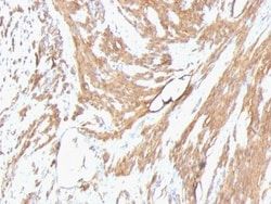
Test Specificity: This antibody recognizes actin of skeletal, cardiac, and smooth muscle cells. It is not reactive with other mesenchymal cells except for myoepithelium. Actin can be resolved on the basis of its isoelectric points into three distinctive components: alpha, beta, and gamma in order of increasing isoelectric point. Anti-muscle specific actin recognizes alpha and gamma isotypes of all muscle groups. Non-muscle cells such as vascular endothelial cells and connective tissues are non-reactive. Also, neoplastic cells of non-muscle-derived tissue such as carcinomas, melanomas, and lymphomas are negative.AIt stains tumors of smooth muscle (leiomyomas and leiomyosarcomas) as well as skeletal muscle (rhabdomyomas and rhabdomyosarcomas).