Actin (Muscle Specific) Antibody (HHF35 + MSA/953), Novus Biologicals™
Mouse Monoclonal Antibody
Manufacturer: Fischer Scientific
The price for this product is unavailable. Please request a quote
Antigen
pan Actin
Concentration
0.2mg/mL
Applications
Flow Cytometry, Immunohistochemistry (Paraffin), Immunofluorescence
Conjugate
Unconjugated
Host Species
Mouse
Research Discipline
Angiogenesis, Cancer, Cell Biology, Cellular Markers, Cytoskeleton Markers, Stem Cell Markers
Formulation
10mM PBS and 0.05% BSA with 0.05% Sodium Azide
Gene Alias
ACTA, actin, alpha 1, skeletal muscle, alpha skeletal muscle, alpha skeletal muscle actin, alpha-actin-1, ASMA, CFTD, CFTDM, MPFD, NEM1, NEM2, NEM3
Gene Symbols
ACTA1
Isotype
IgG1 κ
Purification Method
Protein A or G purified
Test Specificity
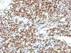
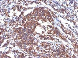
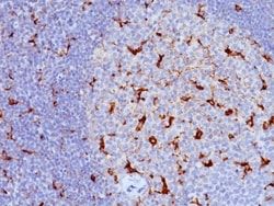
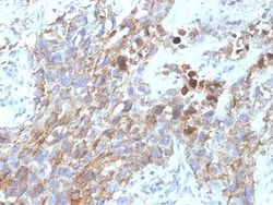
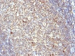
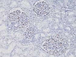
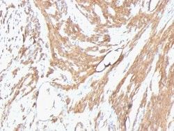
This antibody recognizes actin of skeletal, cardiac, and smooth muscle cells. It is not reactive with other mesenchymal cells except for myoepithelium. Actin can be resolved on the basis of its isoelectric points into three distinctive components: alpha, beta, and gamma in order of increasing isoelectric point. Anti-muscle specific actin recognizes alpha and gamma isotypes of all muscle groups. Non-muscle cells such as vascular endothelial cells and connective tissues are non-reactive. Also, neoplastic cells of non-muscle-derived tissue such as carcinomas, melanomas, and lymphomas are negative.AIt stains tumors of smooth muscle (leiomyomas and leiomyosarcomas) as well as skeletal muscle (rhabdomyomas and rhabdomyosarcomas).
Clone
HHF35 + MSA/953
Dilution
Flow Cytometry 0.5 - 1 ug/million cells in 0.1 ml, Immunohistochemistry-Paraffin 0.25 - 0.5 ug/ml, Immunofluorescence 0.5 - 1.0 ug/ml
Classification
Monoclonal
Form
Purified
Regulatory Status
RUO
Target Species
Human, Rat, Rabbit
Gene Accession No.
P62736, P68032, P68133
Gene ID (Entrez)
58
Immunogen
Recombinant human actin fragment
Primary or Secondary
Primary
Content And Storage
Store at 4C.
Related Products


Description
- Ensure accurate, reproducible results in Flow Cytometry, Immunohistochemistry (Paraffin), Immunofluorescence Actin (Muscle Specific) Monoclonal specifically detects Actin (Muscle Specific) in Human, Rat, Rabbit samples
- It is validated for Flow Cytometry, Immunohistochemistry, Immunocytochemistry/Immunofluorescence, Immunohistochemistry-Paraffin, Immunofluorescence.
Compare Similar Items
Show Difference
Antigen: pan Actin
Concentration: 0.2mg/mL
Applications: Flow Cytometry, Immunohistochemistry (Paraffin), Immunofluorescence
Conjugate: Unconjugated
Host Species: Mouse
Research Discipline: Angiogenesis, Cancer, Cell Biology, Cellular Markers, Cytoskeleton Markers, Stem Cell Markers
Formulation: 10mM PBS and 0.05% BSA with 0.05% Sodium Azide
Gene Alias: ACTA, actin, alpha 1, skeletal muscle, alpha skeletal muscle, alpha skeletal muscle actin, alpha-actin-1, ASMA, CFTD, CFTDM, MPFD, NEM1, NEM2, NEM3
Gene Symbols: ACTA1
Isotype: IgG1 κ
Purification Method: Protein A or G purified
Test Specificity: This antibody recognizes actin of skeletal, cardiac, and smooth muscle cells. It is not reactive with other mesenchymal cells except for myoepithelium. Actin can be resolved on the basis of its isoelectric points into three distinctive components: alpha, beta, and gamma in order of increasing isoelectric point. Anti-muscle specific actin recognizes alpha and gamma isotypes of all muscle groups. Non-muscle cells such as vascular endothelial cells and connective tissues are non-reactive. Also, neoplastic cells of non-muscle-derived tissue such as carcinomas, melanomas, and lymphomas are negative.AIt stains tumors of smooth muscle (leiomyomas and leiomyosarcomas) as well as skeletal muscle (rhabdomyomas and rhabdomyosarcomas).
Clone: HHF35 + MSA/953
Dilution: Flow Cytometry 0.5 - 1 ug/million cells in 0.1 ml, Immunohistochemistry-Paraffin 0.25 - 0.5 ug/ml, Immunofluorescence 0.5 - 1.0 ug/ml
Classification: Monoclonal
Form: Purified
Regulatory Status: RUO
Target Species: Human, Rat, Rabbit
Gene Accession No.: P62736, P68032, P68133
Gene ID (Entrez): 58
Immunogen: Recombinant human actin fragment
Primary or Secondary: Primary
Content And Storage: Store at 4C.
Antigen: Pax6
Concentration: 0.2mg/mL
Applications: Flow Cytometry, Immunohistochemistry (Paraffin), Immunofluorescence
Conjugate: Unconjugated
Host Species: Mouse
Research Discipline: Cellular Markers, Diabetes Research, Neuronal Stem Cell Markers, Neuronal Stem Cells, Neuroscience, Sensory Systems, Stem Cell Markers, Stem Cells, Transcription Factors and Regulators, Vision
Formulation: 10mM PBS and 0.05% BSA with 0.05% Sodium Azide
Gene Alias: keratitis), MGC17209, Oculorhombin, paired box 6, paired box protein Pax-6
Gene Symbols: PAX6
Isotype: IgG1 κ
Purification Method: Protein A or G purified
Test Specificity: Pax genes contain paired domains with strong homology to genes in Drosophila, which are involved in programming early development. Lesions in the Pax-6 gene account for most cases of aniridia, a congenital malformation of the eye, chiefly characterized by iris hypoplasia, which can cause blindness. Pax-6 is involved in other anterior segment malformations besides aniridia, such as Peters anomaly, a major error in the embryonic development of the eye with corneal clouding with variable iridolenticulocorneal adhesions. The Pax-6 gene encodes a transcriptional regulator that recognizes target genes through its paired-type DNA-binding domain. The paired domain is composed of two distinct DNA-binding subdomains, the amino-terminal subdomain and the carboxy-terminal subdomain, which bind respective consensus DNA sequences. The human Pax-6 gene produces two alternatively spliced isoforms that have the distinct structure of the paired domain.
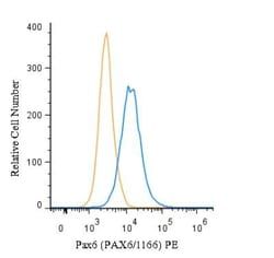
Clone: PAX6/1166
Dilution: Flow Cytometry 0.5 - 1 ug/million cells in 0.1 ml, Immunohistochemistry-Paraffin 0.5 - 1.0 ug/ml, Immunofluorescence 0.5 - 1.0 ug/ml
Classification: Monoclonal
Form: Purified
Regulatory Status: RUO
Target Species: Human
Gene Accession No.: P26367
Gene ID (Entrez): 5080
Immunogen: Recombinant fragment (N-terminus; aa 1-300) of human PAX6 protein
Primary or Secondary: Primary
Content And Storage: Store at 4C.
Antigen: Pax6
Concentration: 0.2mg/mL
Applications: Flow Cytometry, Immunohistochemistry (Paraffin), Immunofluorescence
Conjugate: Unconjugated
Host Species: Mouse
Research Discipline: Cellular Markers, Diabetes Research, Neuronal Stem Cell Markers, Neuronal Stem Cells, Neuroscience, Sensory Systems, Stem Cell Markers, Stem Cells, Transcription Factors and Regulators, Vision
Formulation: 10mM PBS and 0.05% BSA with 0.05% Sodium Azide
Gene Alias: keratitis), MGC17209, Oculorhombin, paired box 6, paired box protein Pax-6
Gene Symbols: PAX6
Isotype: IgG1 κ
Purification Method: Protein A or G purified
Test Specificity: Pax genes contain paired domains with strong homology to genes in Drosophila, which are involved in programming early development. Lesions in the Pax-6 gene account for most cases of aniridia, a congenital malformation of the eye, chiefly characterized by iris hypoplasia, which can cause blindness. Pax-6 is involved in other anterior segment malformations besides aniridia, such as Peters anomaly, a major error in the embryonic development of the eye with corneal clouding with variable iridolenticulocorneal adhesions. The Pax-6 gene encodes a transcriptional regulator that recognizes target genes through its paired-type DNA-binding domain. The paired domain is composed of two distinct DNA-binding subdomains, the amino-terminal subdomain and the carboxy-terminal subdomain, which bind respective consensus DNA sequences. The human Pax-6 gene produces two alternatively spliced isoforms that have the distinct structure of the paired domain.
Clone: PAX6/1166
Dilution: Flow Cytometry 0.5 - 1 ug/million cells in 0.1 ml, Immunohistochemistry-Paraffin 0.5 - 1.0 ug/ml, Immunofluorescence 0.5 - 1.0 ug/ml
Classification: Monoclonal
Form: Purified
Regulatory Status: RUO
Target Species: Human
Gene Accession No.: P26367
Gene ID (Entrez): 5080
Immunogen: Recombinant fragment (N-terminus; aa 1-300) of human PAX6 protein
Primary or Secondary: Primary
Content And Storage: Store at 4C.