Golgi Complex Antibody (AE-6), Novus Biologicals™
Mouse Monoclonal Antibody
Manufacturer: Fischer Scientific
The price for this product is unavailable. Please request a quote
Antigen
Golgi Complex
Concentration
0.2mg/mL
Applications
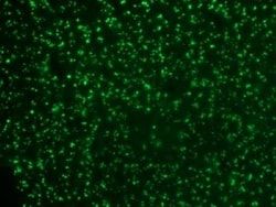
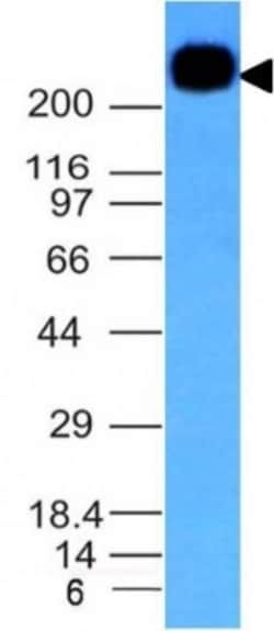
Western Blot, Flow Cytometry, Immunohistochemistry (Paraffin), Immunofluorescence, Immunocytochemistry
Conjugate
Unconjugated
Host Species
Mouse
Target Species
Human, Mouse (Negative), Rat (Negative)
Immunogen
SU-DHL-1 large cell lymphoma cells
Primary or Secondary
Primary
Content And Storage
Store at 4C.
Clone
AE-6
Dilution
Western Blot 0.5 - 1.0 ug/ml, Flow Cytometry 0.5 - 1 ug/million cells in 0.1ml, Immunohistochemistry-Paraffin 0.5 - 1.0 ug/ml, Immunofluorescence 0.5 - 1.0 ug/ml, Immunocytochemistry 0.5 - 1.0 ug/ml
Classification
Monoclonal
Form
Purified
Regulatory Status
RUO
Formulation
10mM PBS and 0.05% BSA with 0.05% Sodium Azide
Isotype
IgG1 κ
Purification Method
Protein A or G purified
Test Specificity
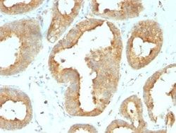
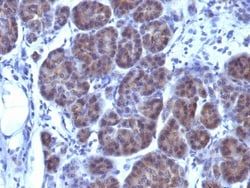
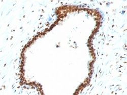
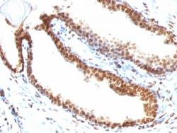
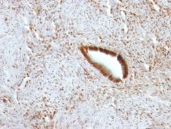
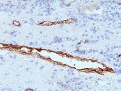
This MAb recognizes an antigen associated with the Golgi complex in human cells only. It can be used to stain the Golgi complex in cell or tissue preparations and can be used as a Golgi marker in subcellular fractions. It produces a diffuse staining pattern of the Golgi zone in normal and malignant cells. This MAb is an excellent marker for human cells in xenographic model research. It reacts specifically with human cells. The Golgi apparatus is an organelle present in all eukaryotic cells that forms a part of the endomembrane system. The primary function of the Golgi apparatus is to process and package macromolecules synthesized by the cell for exocytosis or use within the cell. The Golgi is made up of a stack of flattened, membrane-bound sacs known as cisternae, with three functional regions: the cis face, medial region and trans face. Each region consists of various enzymes that selectively modify the macromolecules passing though them, depending on where they are destined to reside
Related Products
Description
- Ensure accurate, reproducible results in Western Blot, Flow Cytometry, Immunohistochemistry (Paraffin), Immunocytochemistry, Immunofluorescence Golgi Complex Monoclonal specifically detects Golgi Complex in Human, Mouse (Negative), Rat (Negative) samples
- It is validated for Western Blot, Flow Cytometry, Immunohistochemistry, Immunocytochemistry/Immunofluorescence, Immunohistochemistry-Paraffin, Immunofluorescence, Immunocytochemistry.
Compare Similar Items
Show Difference

Antigen: Golgi Complex
Concentration: 0.2mg/mL
Applications: Western Blot, Flow Cytometry, Immunohistochemistry (Paraffin), Immunofluorescence, Immunocytochemistry
Conjugate: Unconjugated
Host Species: Mouse
Target Species: Human, Mouse (Negative), Rat (Negative)
Immunogen: SU-DHL-1 large cell lymphoma cells
Primary or Secondary: Primary
Content And Storage: Store at 4C.
Clone: AE-6
Dilution: Western Blot 0.5 - 1.0 ug/ml, Flow Cytometry 0.5 - 1 ug/million cells in 0.1ml, Immunohistochemistry-Paraffin 0.5 - 1.0 ug/ml, Immunofluorescence 0.5 - 1.0 ug/ml, Immunocytochemistry 0.5 - 1.0 ug/ml
Classification: Monoclonal
Form: Purified
Regulatory Status: RUO
Formulation: 10mM PBS and 0.05% BSA with 0.05% Sodium Azide
Isotype: IgG1 κ
Purification Method: Protein A or G purified
Test Specificity: This MAb recognizes an antigen associated with the Golgi complex in human cells only. It can be used to stain the Golgi complex in cell or tissue preparations and can be used as a Golgi marker in subcellular fractions. It produces a diffuse staining pattern of the Golgi zone in normal and malignant cells. This MAb is an excellent marker for human cells in xenographic model research. It reacts specifically with human cells. The Golgi apparatus is an organelle present in all eukaryotic cells that forms a part of the endomembrane system. The primary function of the Golgi apparatus is to process and package macromolecules synthesized by the cell for exocytosis or use within the cell. The Golgi is made up of a stack of flattened, membrane-bound sacs known as cisternae, with three functional regions: the cis face, medial region and trans face. Each region consists of various enzymes that selectively modify the macromolecules passing though them, depending on where they are destined to reside
Antigen: Golgi Glycoprotein 1/GLG1
Concentration: 0.2mg/mL
Applications: Western Blot, Flow Cytometry, Immunohistochemistry (Paraffin), Immunofluorescence, Immunocytochemistry
Conjugate: Unconjugated
Host Species: Mouse
Target Species: Human, Mouse (Negative), Rat (Negative)
Immunogen: Golgi fraction from human liver cells
Primary or Secondary: Primary
Content And Storage: Store at 4C.
Clone: GLG1/970
Dilution: Western Blot 0.5 - 1.0 ug/ml, Flow Cytometry 0.5 - 1 ug/million cells in 0.1ml, Immunohistochemistry-Paraffin 0.5 - 1.0 ug/ml, Immunofluorescence 0.5 - 1.0 ug/ml, Immunocytochemistry 0.5 - 1.0 ug/ml
Classification: Monoclonal
Form: Purified
Regulatory Status: RUO
Formulation: 10mM PBS and 0.05% BSA with 0.05% Sodium Azide
Isotype: IgG1 κ
Purification Method: Protein A or G purified
Test Specificity: This MAb recognizes a protein of 134kDa, which binds fibroblast growth factor and E-selectin (cell-adhesion lectin on endothelial cells mediating the binding of neutrophils). Fucosylation is essential for binding to E-selectin. It contains sialic acid residues and 16 Cys-rich GLG1 repeats. This MAb can be used to stain the Golgi complex in cell or tissue preparations and can be used as a Golgi marker in subcellular fractions. It produces a diffuse staining pattern of the Golgi zone in normal and malignant cells. This MAb is an excellent marker for human cells in xenographic model research. It reacts specifically with human cells. The Golgi apparatus is an organelle present in all eukaryotic cells that forms a part of the endomembrane system. The primary function of the Golgi apparatus is to process and package macromolecules synthesized by the cell for exocytosis or use within the cell. The Golgi is made up of a stack of flattened, membrane-bound sacs known as cisternae, with three fun
Antigen: __
Concentration: __
Applications: __
Conjugate: __
Host Species: __
Target Species: __
Immunogen: __
Primary or Secondary: __
Content And Storage: __
Clone: __
Dilution: __
Classification: __
Form: __
Regulatory Status: __
Formulation: __
Isotype: __
Purification Method: __
Test Specificity: __