Von Willebrand Factor Antibody (IIIE2.34), Novus Biologicals™
Mouse Monoclonal Antibody
Manufacturer: Fischer Scientific
The price for this product is unavailable. Please request a quote
Antigen
Von Willebrand Factor
Concentration
0.2 mg/mL
Applications
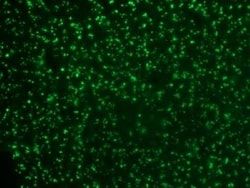
Western Blot, Flow Cytometry, Immunoprecipitation, Immunohistochemistry (Paraffin), Immunofluorescence
Conjugate
Unconjugated
Host Species
Mouse
Research Discipline
Cancer
Formulation
10mM PBS and 0.05% BSA with 0.05% Sodium Azide
Gene Alias
coagulation factor VIII VWF, F8, F8VWF, von Willebrand factor, VWD, vWF
Gene Symbols
VWF
Isotype
IgG1 κ
Purification Method
Protein A or G purified
Test Specificity
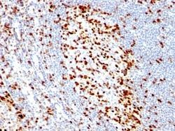
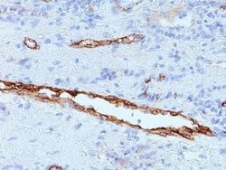
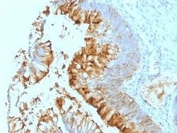
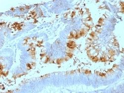
Von Willebrand Factor (vWF) is a multimeric glycoprotein that is found in endothelial cells, plasma and platelets. It acts as a carrier protein for Factor VIII and promotes platelet adhesion and aggregation. vWF undergoes a variety of posttranslational modifications that influence the affinity and availability for Factor VIII, including cleavage of the propeptide and formation of N-terminal disulfide bonds. This antibody helps to establish the endothelial nature of some lesions of disputed histogenesis, e.g. Kaposi's sarcoma and cardiac myxoma. It is widely used for differentiating vascular lesions from those of other tissue differentiation within a panel of other vascular markers although not all tumors of endothelial differentiation contain this antigen.
Clone
IIIE2.34
Dilution
Western Blot 0.5 - 1.0 ug/ml, Flow Cytometry 0.5 - 1 ug/million cells in 0.1 ml, Immunoprecipitation 0.5 - 1 ug/500 ug Protein Lysate, Immunohistochemistry-Paraffin 0.5 - 1.0 ug/ml, Immunofluorescence 0.5 - 1.0 ug/ml
Classification
Monoclonal
Form
Purified
Regulatory Status
RUO
Target Species
Human
Gene Accession No.
P04275
Gene ID (Entrez)
7450
Immunogen
Recombinant human vWF fragment spanning aa 845-949
Primary or Secondary
Primary
Content And Storage
Store at 4C.
Molecular Weight of Antigen
250 kDa
Related Products


Description
- Ensure accurate, reproducible results in Western Blot, Flow Cytometry, Immunohistochemistry (Paraffin), Immunoprecipitation, Immunofluorescence Von Willebrand Factor Monoclonal specifically detects Von Willebrand Factor in Human samples
- It is validated for Western Blot, Flow Cytometry, Immunohistochemistry, Immunocytochemistry/Immunofluorescence, Immunoprecipitation, Immunohistochemistry-Paraffin, Immunofluorescence.
Compare Similar Items
Show Difference
Antigen: Von Willebrand Factor
Concentration: 0.2 mg/mL
Applications: Western Blot, Flow Cytometry, Immunoprecipitation, Immunohistochemistry (Paraffin), Immunofluorescence
Conjugate: Unconjugated
Host Species: Mouse
Research Discipline: Cancer
Formulation: 10mM PBS and 0.05% BSA with 0.05% Sodium Azide
Gene Alias: coagulation factor VIII VWF, F8, F8VWF, von Willebrand factor, VWD, vWF
Gene Symbols: VWF
Isotype: IgG1 κ
Purification Method: Protein A or G purified
Test Specificity: Von Willebrand Factor (vWF) is a multimeric glycoprotein that is found in endothelial cells, plasma and platelets. It acts as a carrier protein for Factor VIII and promotes platelet adhesion and aggregation. vWF undergoes a variety of posttranslational modifications that influence the affinity and availability for Factor VIII, including cleavage of the propeptide and formation of N-terminal disulfide bonds. This antibody helps to establish the endothelial nature of some lesions of disputed histogenesis, e.g. Kaposi's sarcoma and cardiac myxoma. It is widely used for differentiating vascular lesions from those of other tissue differentiation within a panel of other vascular markers although not all tumors of endothelial differentiation contain this antigen.
Clone: IIIE2.34
Dilution: Western Blot 0.5 - 1.0 ug/ml, Flow Cytometry 0.5 - 1 ug/million cells in 0.1 ml, Immunoprecipitation 0.5 - 1 ug/500 ug Protein Lysate, Immunohistochemistry-Paraffin 0.5 - 1.0 ug/ml, Immunofluorescence 0.5 - 1.0 ug/ml
Classification: Monoclonal
Form: Purified
Regulatory Status: RUO
Target Species: Human
Gene Accession No.: P04275
Gene ID (Entrez): 7450
Immunogen: Recombinant human vWF fragment spanning aa 845-949
Primary or Secondary: Primary
Content And Storage: Store at 4C.
Molecular Weight of Antigen: 250 kDa
Antigen: WT1
Concentration: 0.2mg/mL
Applications: Flow Cytometry, Immunohistochemistry (Paraffin), Immunofluorescence
Conjugate: Unconjugated
Host Species: Mouse
Research Discipline: Angiogenesis, Cancer, Cell Cycle and Replication, Cellular Markers, Chromatin Research, Tumor Suppressors
Formulation: 1.0mM PBS and 0.05% BSA with 0.05% Sodium Azide
Gene Alias: AWT1, NPHS4GUD, WAGR, Wilms tumor 1, Wilms tumor protein, WIT-2, WT33
Gene Symbols: WT1
Isotype: IgG1 κ
Purification Method: Protein A or G purified
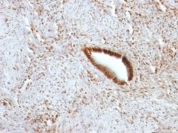
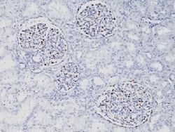
Test Specificity: Recognizes a 47-55kDa-tumor suppressor protein, identified as Wilm's Tumor (WT1) protein. The antibody reacts with all isoforms of the full-length WT1 and also identifies WT1 lacking exon 2-encoded amino acids, frequently found in subsets of sporadic Wilm s tumors.WT1, a sporadic and familial pediatric kidney tumor, is genetically heterogeneous. Wilm s tumor is associated with mutations of WT1, a zinc-finger transcription factor that is essential for the development of the metanephric kidney and the urogenital system. The WT1 gene is normally expressed in fetal kidney and mesothelium, and its expression has been suggested as a marker for Wilm s tumor and mesothelioma. WT1 protein has been identified in proliferative mesothelial cells, malignant mesothelioma, ovarian carcinoma, gonadoblastoma, nephroblastoma, and desmoplastic small round cell tumor. Lung adenocarcinomas rarely stain positive with this antibody. WT1 protein expression in mesothelial cells has become a reliable marker for
Clone: WT1/857
Dilution: Flow Cytometry 0.5 - 1 ug/million cells in 0.1 ml, Immunohistochemistry-Paraffin 0.5 - 1.0 ug/ml, Immunofluorescence 0.5 - 1.0 ug/ml
Classification: Monoclonal
Form: Purified
Regulatory Status: RUO
Target Species: Human, Mouse, Rat
Gene Accession No.: P19544
Gene ID (Entrez): 7490
Immunogen: Recombinant human WT1 protein
Primary or Secondary: Primary
Content And Storage: Store at 4C.
Molecular Weight of Antigen: __
Antigen: ZAP70
Concentration: 0.2mg/mL
Applications: Flow Cytometry, Immunohistochemistry (Paraffin), Immunofluorescence
Conjugate: Unconjugated
Host Species: Mouse
Research Discipline: Adaptive Immunity, Cell Biology, Immunology, Protein Kinase, Signal Transduction, Tyrosine Kinases
Formulation: 1.0mM PBS and 0.05% BSA with 0.05% Sodium Azide
Gene Alias: 70 kDa zeta-associated protein, EC 2.7.10, EC 2.7.10.2, SRKFLJ17679, STDFLJ17670, Syk-related tyrosine kinase, tyrosine-protein kinase ZAP-70, TZK, ZAP-70, zeta-chain (TCR) associated protein kinase (70 kD), zeta-chain (TCR) associated protein kinase 70kDa, zeta-chain associated protein kinase, 70kD
Gene Symbols: ZAP70
Isotype: IgG2a κ
Purification Method: Protein A or G purified
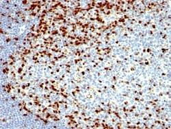
Test Specificity: ZAP70 is a 70kDa protein tyrosine kinase found in T-cells and natural killer cells. Control of this protein translation is via the IgVH gene. In Western blotting of whole cell lysates of normal peripheral blood mononuclear cells, the antibody labels a band corresponding to ZAP70. In Western blotting of whole cell lysates of CD19-positive purified leukemia cells from patients with Ig-unmutated and Ig-mutated CLL, the antibody labels a band corresponding to ZAP70 in the Ig-unmutated CLL samples, whereas no band is observed in the Ig-mutated CLL samples. In Western blotting of cell lysates of Jurkat cells (T-lymphoblastic cell line), the antibody labels a band of 70kDa protein. In Western blotting of cell lysates of A431 cells (carcinoma cell line), no band is observed. ZAP70 protein is expressed in leukemic cells of approximately 25% of chronic lymphocytic leukemia (CLL) cases as well. Anti-ZAP70 expression is an excellent surrogate marker for the distinction between the Ig-mutated (anti
Clone: SPM362
Dilution: Flow Cytometry 0.5 - 1 ug/million cells in 0.1 ml, Immunohistochemistry-Paraffin 0.5 - 1.0 ug/ml, Immunofluorescence 0.5 - 1.0 ug/ml
Classification: Monoclonal
Form: Purified
Regulatory Status: RUO
Target Species: Human
Gene Accession No.: __
Gene ID (Entrez): 7535
Immunogen: Recombinant ZAP-70 protein including residues 1-254 and encompassing SH2 domains of human ZAP70
Primary or Secondary: Primary
Content And Storage: Store at 4C.
Molecular Weight of Antigen: 70 kDa