DNA Antibody (DSD/958), Novus Biologicals™
Mouse Monoclonal Antibody
Manufacturer: Fischer Scientific
The price for this product is unavailable. Please request a quote
Antigen
DNA
Concentration
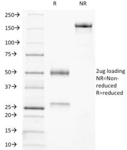
0.2mg/mL
Applications
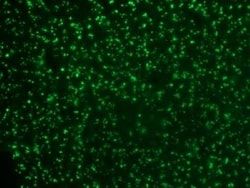
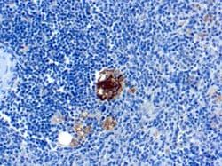
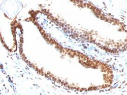
Flow Cytometry, Immunohistochemistry (Paraffin), Immunofluorescence, Immunocytochemistry
Conjugate
Unconjugated
Host Species
Mouse
Target Species
Human
Gene Alias
Double stranded deoxyribonucleic acid, Double stranded DNA, dsDNA
Isotype
IgG3 κ
Purification Method
Protein A or G purified
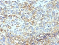
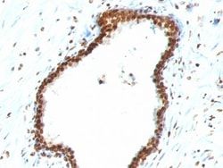
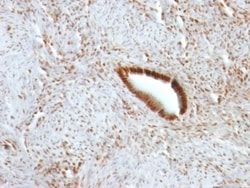
Test Specificity
This MAb recognizes the double stranded DNA in human cells. It can be used to stain the nuclei in cell or tissue preparations and can be used as a nuclear marker in human cells. This MAb produces a homogeneous staining pattern in the nucleus of normal and malignant cells. Deoxyribonucleic acid (DNA) is a nucleic acid that stores long-term information regarding the development and function of all known living organisms. DNA consists of two long nucleotide polymers, which are composed of four bases, namely adenine, thymine, guanine and cytosine, all of which are flanked by a phosphate-deoxyribose backbone. Normally, DNA exists as a double-stranded (ds) molecule that forms in the shape of a double helix, allowing the bases and the backbone of the two strands to interact, thus forming a polynucleotide. When the double helix is unwound (either by enzymes or heat), DNA exists as a single-stranded (ss) molecule that is less stable than the double helix, but is necessary for protein access to
Clone
DSD/958
Dilution
Flow Cytometry 0.5 - 1 ug/million cells in 0.1 ml, Immunohistochemistry-Paraffin 1 - 2 ug/ml, Immunofluorescence 1 - 2 ug/ml, Immunocytochemistry 0.5 - 1.0 ug/ml
Classification
Monoclonal
Form
Purified
Regulatory Status
RUO
Formulation
10mM PBS and 0.05% BSA with 0.05% Sodium Azide
Immunogen
Nuclei of Burkitt's cells
Primary or Secondary
Primary
Content And Storage
Store at 4C.
Related Products

Description
- Ensure accurate, reproducible results in Flow Cytometry, Immunohistochemistry (Paraffin), Immunocytochemistry, Immunofluorescence DNA Monoclonal specifically detects DNA in Human samples
- It is validated for Immunohistochemistry, Immunohistochemistry-Paraffin.
Compare Similar Items
Show Difference
Antigen: DNA
Concentration: 0.2mg/mL
Applications: Flow Cytometry, Immunohistochemistry (Paraffin), Immunofluorescence, Immunocytochemistry
Conjugate: Unconjugated
Host Species: Mouse
Target Species: Human
Gene Alias: Double stranded deoxyribonucleic acid, Double stranded DNA, dsDNA
Isotype: IgG3 κ
Purification Method: Protein A or G purified
Test Specificity: This MAb recognizes the double stranded DNA in human cells. It can be used to stain the nuclei in cell or tissue preparations and can be used as a nuclear marker in human cells. This MAb produces a homogeneous staining pattern in the nucleus of normal and malignant cells. Deoxyribonucleic acid (DNA) is a nucleic acid that stores long-term information regarding the development and function of all known living organisms. DNA consists of two long nucleotide polymers, which are composed of four bases, namely adenine, thymine, guanine and cytosine, all of which are flanked by a phosphate-deoxyribose backbone. Normally, DNA exists as a double-stranded (ds) molecule that forms in the shape of a double helix, allowing the bases and the backbone of the two strands to interact, thus forming a polynucleotide. When the double helix is unwound (either by enzymes or heat), DNA exists as a single-stranded (ss) molecule that is less stable than the double helix, but is necessary for protein access to
Clone: DSD/958
Dilution: Flow Cytometry 0.5 - 1 ug/million cells in 0.1 ml, Immunohistochemistry-Paraffin 1 - 2 ug/ml, Immunofluorescence 1 - 2 ug/ml, Immunocytochemistry 0.5 - 1.0 ug/ml
Classification: Monoclonal
Form: Purified
Regulatory Status: RUO
Formulation: 10mM PBS and 0.05% BSA with 0.05% Sodium Azide
Immunogen: Nuclei of Burkitt's cells
Primary or Secondary: Primary
Content And Storage: Store at 4C.

Antigen: DPPIV/CD26
Concentration: 0.2 mg/mL
Applications: Flow Cytometry, Immunohistochemistry (Frozen), Immunofluorescence
Conjugate: Unconjugated
Host Species: Mouse
Target Species: Human
Gene Alias: ADABP, ADCP-2, ADCP2DPP IV, Adenosine deaminase complexing protein 2TP103, CD26 antigen, CD26T-cell activation antigen CD26, dipeptidyl peptidase 4, Dipeptidyl peptidase IV, dipeptidylpeptidase 4, dipeptidyl-peptidase 4, dipeptidylpeptidase IV (CD26, adenosine deaminase complexing protein 2), DPPIV, EC 3.4.14.5
Isotype: IgM κ
Purification Method: Protein A or G purified
Test Specificity: Recognizes a glycoprotein of 110kDa, identified as CD26. It is an atypical serine protease belonging to the prolyl oligopeptidase family. It is expressed on lymphocyte cells and is upregulated during T-cell activation. CD26 is also expressed on activated B cells and natural killer cells and abundantly on epithelia. CD26 is implicated in a variety of biological functions including T-cell activation, cell adhesion with extracellular matrix such as fibronectin or collagens, and in HIV infection.
Clone: 134-2C2
Dilution: Flow Cytometry 0.5 - 1 ug/million cells in 0.1 ml, Immunohistochemistry-Frozen 0.5 - 1.0 ug/ml, Immunofluorescence 0.5 - 1.0 ug/ml
Classification: Monoclonal
Form: Purified
Regulatory Status: RUO
Formulation: 10mM PBS and 0.05% BSA with 0.05% Sodium Azide
Immunogen: A synthetic peptide from the amino terminal region of CD26.
Primary or Secondary: Primary
Content And Storage: Store at 4C.
Antigen: DPPIV/CD26
Concentration: 0.2 mg/mL
Applications: Flow Cytometry, Immunohistochemistry (Frozen), Immunofluorescence
Conjugate: Unconjugated
Host Species: Mouse
Target Species: Human, Rat, Porcine (Negative), Sheep (Negative)
Gene Alias: ADABP, ADCP-2, ADCP2DPP IV, Adenosine deaminase complexing protein 2TP103, CD26 antigen, CD26T-cell activation antigen CD26, dipeptidyl peptidase 4, Dipeptidyl peptidase IV, dipeptidylpeptidase 4, dipeptidyl-peptidase 4, dipeptidylpeptidase IV (CD26, adenosine deaminase complexing protein 2), DPPIV, EC 3.4.14.5
Isotype: IgG2b κ
Purification Method: Protein A or G purified
Test Specificity: Recognizes a glycoprotein of 110kDa, identified as CD26 (Workshop VI; Code: N-L039). It is an atypical serine protease belonging to the prolyl oligopeptidase family. It is expressed on lymphocyte cells and is upregulated during T-cell activation. CD26 is also expressed on activated B cells and natural killer cells and abundantly on epithelia. CD26 is implicated in a variety of biological functions including T-cell activation, cell adhesion with extracellular matrix such as fibronectin or collagens, and in HIV infection. Cross-linking of CD26 using this antibody dramatically enhances the anti-CD3-induced IL-2 production. In Western blotting, this MAb reacts with only glycosylated CD26, but not with the deglycosylated form. It does not prevent ADA binding to CD26.
Clone: 202.36
Dilution: Flow Cytometry 0.5 - 1 ug/million cells in 0.1 ml, Immunohistochemistry-Frozen 0.5 - 1.0 ug/ml, Immunofluorescence 0.5 - 1.0 ug/ml
Classification: Monoclonal
Form: Purified
Regulatory Status: RUO
Formulation: 10mM PBS and 0.05% BSA with 0.05% Sodium Azide
Immunogen: Human T cell clone
Primary or Secondary: Primary
Content And Storage: Store at 4C.