EpCAM/TROP1 Antibody (SPM491) - (ECD), Novus Biologicals™
Mouse Monoclonal Antibody
Manufacturer: Fischer Scientific
The price for this product is unavailable. Please request a quote
Antigen
EpCAM/TROP1
Concentration
0.2mg/mL
Applications
Western Blot, Flow Cytometry, Immunohistochemistry (Paraffin), Immunofluorescence
Conjugate
Unconjugated
Host Species
Mouse
Research Discipline
Cancer, Cancer Stem Cells
Formulation
10mM PBS and 0.05% BSA with 0.05% Sodium Azide
Gene ID (Entrez)
4072
Immunogen
Recombinant full-length human EpCAM protein
Primary or Secondary
Primary
Content And Storage
Store at 4C.
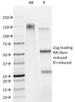
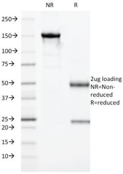
Molecular Weight of Antigen
41 kDa
Clone
SPM491
Dilution
Western Blot 0.5 - 1.0 ug/ml, Flow Cytometry 0.5 - 1 ug/million cells in 0.1 ml, Immunohistochemistry-Paraffin 0.25 - 0.5 ug/ml, Immunofluorescence 1 - 2 ug/ml
Classification
Monoclonal
Form
Purified
Regulatory Status
RUO
Target Species
Human, Mouse (Negative), Rat (Negative)
Gene Alias
17-1A, 323/A3, ACSTD1, antigen identified by monoclonal AUA1, CD326 antigen, Cell surface glycoprotein Trop-1, chromosome 4, surface marker (35kD glycoprotein), DIAR5, EGP, EGP-2, EGP314, EGP40, EpCAM, epithelial cell adhesion molecule, Epithelial cell surface antigen, Epithelial glycoprotein, Epithelial glycoprotein 314, ESA, GA733-2EGP34, hEGP314, HNPCC8, KS 1/4 antigen, KS1/4, KSAHEA125, M1S2, M4S1Ly74, Major gastrointestinal tumor-associated protein GA733-2, MIC18MH99, MOC31, TACST-1, TACSTD1, TROP1CD326, Tumor-associated calcium signal transducer 1CO-17A
Gene Symbols
EPCAM
Isotype
IgG1 κ
Purification Method
Protein A or G purified
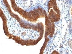
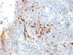
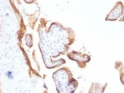
Test Specificity
EGP40 is a 40-43kDa transmembrane epithelial glycoprotein, also identified as epithelial specific antigen (ESA), or epithelial cellular adhesion molecule (Ep-CAM). It is expressed on baso-lateral cell surface in most simple epithelia and a vast majority of carcinomas. This antibody has been used to distinguish adenocarcinoma from pleural mesothelioma and hepatocellular carcinoma. This antibody is also useful in distinguishing serous carcinomas of the ovary from mesothelioma.
Related Products

Description
- Ensure accurate, reproducible results in Western Blot, Flow Cytometry, Immunohistochemistry (Paraffin), Immunofluorescence EpCAM/TROP1 Monoclonal specifically detects EpCAM/TROP1 in Human, Mouse (Negative), Rat (Negative) samples
- It is validated for Western Blot, Flow Cytometry, Immunohistochemistry, Immunocytochemistry/Immunofluorescence, Immunohistochemistry-Paraffin, Immunofluorescence.
Compare Similar Items
Show Difference
Antigen: EpCAM/TROP1
Concentration: 0.2mg/mL
Applications: Western Blot, Flow Cytometry, Immunohistochemistry (Paraffin), Immunofluorescence
Conjugate: Unconjugated
Host Species: Mouse
Research Discipline: Cancer, Cancer Stem Cells
Formulation: 10mM PBS and 0.05% BSA with 0.05% Sodium Azide
Gene ID (Entrez): 4072
Immunogen: Recombinant full-length human EpCAM protein
Primary or Secondary: Primary
Content And Storage: Store at 4C.
Molecular Weight of Antigen: 41 kDa
Clone: SPM491
Dilution: Western Blot 0.5 - 1.0 ug/ml, Flow Cytometry 0.5 - 1 ug/million cells in 0.1 ml, Immunohistochemistry-Paraffin 0.25 - 0.5 ug/ml, Immunofluorescence 1 - 2 ug/ml
Classification: Monoclonal
Form: Purified
Regulatory Status: RUO
Target Species: Human, Mouse (Negative), Rat (Negative)
Gene Alias: 17-1A, 323/A3, ACSTD1, antigen identified by monoclonal AUA1, CD326 antigen, Cell surface glycoprotein Trop-1, chromosome 4, surface marker (35kD glycoprotein), DIAR5, EGP, EGP-2, EGP314, EGP40, EpCAM, epithelial cell adhesion molecule, Epithelial cell surface antigen, Epithelial glycoprotein, Epithelial glycoprotein 314, ESA, GA733-2EGP34, hEGP314, HNPCC8, KS 1/4 antigen, KS1/4, KSAHEA125, M1S2, M4S1Ly74, Major gastrointestinal tumor-associated protein GA733-2, MIC18MH99, MOC31, TACST-1, TACSTD1, TROP1CD326, Tumor-associated calcium signal transducer 1CO-17A
Gene Symbols: EPCAM
Isotype: IgG1 κ
Purification Method: Protein A or G purified
Test Specificity: EGP40 is a 40-43kDa transmembrane epithelial glycoprotein, also identified as epithelial specific antigen (ESA), or epithelial cellular adhesion molecule (Ep-CAM). It is expressed on baso-lateral cell surface in most simple epithelia and a vast majority of carcinomas. This antibody has been used to distinguish adenocarcinoma from pleural mesothelioma and hepatocellular carcinoma. This antibody is also useful in distinguishing serous carcinomas of the ovary from mesothelioma.
Antigen: ER beta/NR3A2
Concentration: 0.2mg/mL
Applications: Western Blot, Flow Cytometry, Immunohistochemistry (Paraffin), SDS-Page, Immunofluorescence
Conjugate: Unconjugated
Host Species: Mouse
Research Discipline: Breast Cancer, Cancer, Cell Biology, Cell Cycle and Replication, Dendritic Cell Markers, GPCR, Neuroscience, Signal Transduction, Transcription Factors and Regulators
Formulation: 10mM PBS and 0.05% BSA with 0.05% Sodium Azide
Gene ID (Entrez): 2100
Immunogen: C-terminus fragment of recombinant human estrogen receptor beta protein
Primary or Secondary: Primary
Content And Storage: Store at 4C.
Molecular Weight of Antigen: __
Clone: ESR2/686
Dilution: Western Blot 0.5 - 1.0 ug/ml, Flow Cytometry 0.5 - 1 ug/million cells in 0.1 ml, Immunohistochemistry-Paraffin 0.5 - 1.0 ug/ml, SDS-Page, Immunofluorescence 0.5 - 1.0 ug/ml
Classification: Monoclonal
Form: Purified
Regulatory Status: RUO
Target Species: Human
Gene Alias: Erb, ER-beta, ESR-BETA, ESTRB, estrogen receptor 2 (ER beta), estrogen receptor beta, estrogen receptor beta 4, NR3A2ESRB, Nuclear receptor subfamily 3 group A member 2
Gene Symbols: ESR2
Isotype: IgG2a
Purification Method: Protein A or G purified
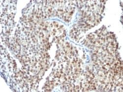
Test Specificity: Estrogen receptors (ER) are members of the steroid/thyroid hormone receptor superfamily of ligand-activated transcription factors. Estrogen receptors, including ER-alpha and ER-beta, contain DNA binding and ligand binding domains and are critically involved in regulating the normal function of reproductive tissues. They are located in the nucleus, though some estrogen receptors associate with the cell surface membrane and can be rapidly activated by exposure of cells to estrogen. ER-alpha and ER-beta are differentially activated by various ligands. Receptor-ligand interactions trigger a cascade of events, including dissociation from heat shock proteins, receptor dimerization, phosphorylation and the association of the hormone activated receptor with specific regulatory elements in target genes. Evidence suggests that ER-alpha and ER-beta may be regulated by distinct mechanisms even though they share many functional characteristics.
Antigen: ER beta/NR3A2
Concentration: 0.2mg/mL
Applications: Western Blot, Flow Cytometry, Immunohistochemistry (Paraffin), SDS-Page, Immunofluorescence
Conjugate: Unconjugated
Host Species: Mouse
Research Discipline: Breast Cancer, Cancer, Cell Biology, Cell Cycle and Replication, Dendritic Cell Markers, GPCR, Neuroscience, Signal Transduction, Transcription Factors and Regulators
Formulation: 10mM PBS and 0.05% BSA with 0.05% Sodium Azide
Gene ID (Entrez): 2100
Immunogen: C-terminus fragment of recombinant human estrogen receptor beta protein
Primary or Secondary: Primary
Content And Storage: Store at 4C.
Molecular Weight of Antigen: __
Clone: ESR2/686
Dilution: Western Blot 0.5 - 1.0 ug/ml, Flow Cytometry 0.5 - 1 ug/million cells in 0.1 ml, Immunohistochemistry-Paraffin 0.5 - 1.0 ug/ml, SDS-Page, Immunofluorescence 0.5 - 1.0 ug/ml
Classification: Monoclonal
Form: Purified
Regulatory Status: RUO
Target Species: Human
Gene Alias: Erb, ER-beta, ESR-BETA, ESTRB, estrogen receptor 2 (ER beta), estrogen receptor beta, estrogen receptor beta 4, NR3A2ESRB, Nuclear receptor subfamily 3 group A member 2
Gene Symbols: ESR2
Isotype: IgG2a
Purification Method: Protein A or G purified
Test Specificity: Estrogen receptors (ER) are members of the steroid/thyroid hormone receptor superfamily of ligand-activated transcription factors. Estrogen receptors, including ER-alpha and ER-beta, contain DNA binding and ligand binding domains and are critically involved in regulating the normal function of reproductive tissues. They are located in the nucleus, though some estrogen receptors associate with the cell surface membrane and can be rapidly activated by exposure of cells to estrogen. ER-alpha and ER-beta are differentially activated by various ligands. Receptor-ligand interactions trigger a cascade of events, including dissociation from heat shock proteins, receptor dimerization, phosphorylation and the association of the hormone activated receptor with specific regulatory elements in target genes. Evidence suggests that ER-alpha and ER-beta may be regulated by distinct mechanisms even though they share many functional characteristics.