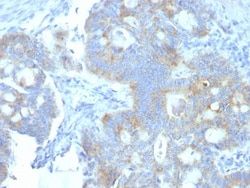
EGFR Antibody (SPM622), Novus Biologicals™
Mouse Monoclonal Antibody
Manufacturer: Fischer Scientific
The price for this product is unavailable. Please request a quote
Antigen
EGFR
Concentration
0.2mg/mL
Applications
Flow Cytometry, Immunohistochemistry (Paraffin), Immunofluorescence
Conjugate
Unconjugated
Host Species
Mouse
Research Discipline
Cancer, Cell Biology, Cell Cycle and Replication, Growth and Development, Hypoxia, Signal Transduction, Tumor Suppressors, Tyrosine Kinases
Formulation
10mM PBS and 0.05% BSA with 0.05% Sodium Azide
Gene Alias
avian erythroblastic leukemia viral (v-erb-b) oncogene homolog, cell growth inhibiting protein 40, cell proliferation-inducing protein 61, EC 2.7.10, EC 2.7.10.1, epidermal growth factor receptor, epidermal growth factor receptor (avian erythroblastic leukemia viral (v-erb-b)oncogene homolog), ERBB, ErbB1, ERBB1PIG61, HER1, mENA, Proto-oncogene c-ErbB-1, Receptor tyrosine-protein kinase erbB-1
Gene Symbols
EGFR
Isotype
IgG1 κ
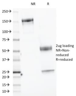
Purification Method
Protein A or G purified
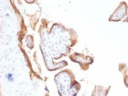
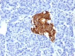
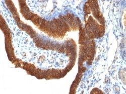
Test Specificity
This MAb recognizes a protein of 170kDa, identified as EGFR. EGFR is type I receptor tyrosine kinase with sequence homology to erbB-1, -2, -3 -4 or HER-1, -2, -3 -4. It binds to Epidermal Growth Factor (EGF), Transforming Growth Factor-a (TGF-a), Heparin-binding EGF (HB-EGF), amphiregulin, Beta cellulin and epiregulin. EGFR is overexpressed in tumors of breast, brain, bladder, lung, gastric, head & neck, esophagus, cervix, vulva, ovary, and endometrium. It is predominantly present in squamous cell carcinomas.
Clone
SPM622
Dilution
Flow Cytometry 0.5 - 1 ug/million cells in 0.1 ml, Immunohistochemistry-Paraffin 2 - 4 ug/ml, Immunofluorescence 1 - 2 ug/ml
Classification
Monoclonal
Form
Purified
Regulatory Status
RUO
Target Species
Human
Gene Accession No.
P00533
Gene ID (Entrez)
1956
Immunogen
Human EGFR purified from A431 cells
Primary or Secondary
Primary
Content And Storage
Store at 4C.
Related Products

Description
- Ensure accurate, reproducible results in Flow Cytometry, Immunohistochemistry (Paraffin), Immunofluorescence EGFR Monoclonal specifically detects EGFR in Human samples
- It is validated for Flow Cytometry, Immunohistochemistry, Immunocytochemistry/Immunofluorescence, Immunohistochemistry-Paraffin, Immunofluorescence.
Compare Similar Items
Show Difference
Antigen: EGFR
Concentration: 0.2mg/mL
Applications: Flow Cytometry, Immunohistochemistry (Paraffin), Immunofluorescence
Conjugate: Unconjugated
Host Species: Mouse
Research Discipline: Cancer, Cell Biology, Cell Cycle and Replication, Growth and Development, Hypoxia, Signal Transduction, Tumor Suppressors, Tyrosine Kinases
Formulation: 10mM PBS and 0.05% BSA with 0.05% Sodium Azide
Gene Alias: avian erythroblastic leukemia viral (v-erb-b) oncogene homolog, cell growth inhibiting protein 40, cell proliferation-inducing protein 61, EC 2.7.10, EC 2.7.10.1, epidermal growth factor receptor, epidermal growth factor receptor (avian erythroblastic leukemia viral (v-erb-b)oncogene homolog), ERBB, ErbB1, ERBB1PIG61, HER1, mENA, Proto-oncogene c-ErbB-1, Receptor tyrosine-protein kinase erbB-1
Gene Symbols: EGFR
Isotype: IgG1 κ
Purification Method: Protein A or G purified
Test Specificity: This MAb recognizes a protein of 170kDa, identified as EGFR. EGFR is type I receptor tyrosine kinase with sequence homology to erbB-1, -2, -3 -4 or HER-1, -2, -3 -4. It binds to Epidermal Growth Factor (EGF), Transforming Growth Factor-a (TGF-a), Heparin-binding EGF (HB-EGF), amphiregulin, Beta cellulin and epiregulin. EGFR is overexpressed in tumors of breast, brain, bladder, lung, gastric, head & neck, esophagus, cervix, vulva, ovary, and endometrium. It is predominantly present in squamous cell carcinomas.
Clone: SPM622
Dilution: Flow Cytometry 0.5 - 1 ug/million cells in 0.1 ml, Immunohistochemistry-Paraffin 2 - 4 ug/ml, Immunofluorescence 1 - 2 ug/ml
Classification: Monoclonal
Form: Purified
Regulatory Status: RUO
Target Species: Human
Gene Accession No.: P00533
Gene ID (Entrez): 1956
Immunogen: Human EGFR purified from A431 cells
Primary or Secondary: Primary
Content And Storage: Store at 4C.
Antigen: Elastase
Concentration: 0.2 mg/mL
Applications: Western Blot, Flow Cytometry, SDS-Page, Immunofluorescence
Conjugate: Unconjugated
Host Species: Mouse
Research Discipline: Cancer, Neuroscience
Formulation: 10mM PBS and 0.05% BSA with 0.05% Sodium Azide
Gene Alias: CBPP, cholesterol-binding pancreatic protease, chymotrypsin-like elastase family member 3B, chymotrypsin-like elastase family, member 3B, E1, EC 3.4.21, EC 3.4.21.70, EL-1, ELA3B, elastase 1, elastase 3B, pancreatic, Elastase IIIB, elastase-3B, fecal elastase 1, pancreatic elastase 1, pancreatic endopeptidase E, Protease E, proteinase E
Gene Symbols: CELA3B
Isotype: IgG κ
Purification Method: Protein A or G purified
Test Specificity: This MAb recognizes a protein of ∼17kDa, identified as CELA3B (Chymotrypsin like elastase family member 3B). Elastases form a subfamily of serine proteases that hydrolyze many proteins in addition to elastin. Humans have six elastase genes which encode the structurally similar proteins elastase 1, 2, 2A, 2B, 3A, and 3B. Unlike other elastases, elastase 3B has little elastolytic activity. Like most of the human elastases, elastase 3B is secreted from the pancreas as a zymogen and, like other serine proteases such as trypsin, chymotrypsin and kallikrein; it has a digestive function in the intestine. Elastase 3B preferentially cleaves proteins after alanine residues. Elastase 3B may also function in the intestinal transport and metabolism of cholesterol. Both elastase 3A and elastase 3B have been referred to as protease E and as elastase 1, and excretion of this protein in fecal material is frequently used as a measure of pancreatic function in clinical assays.
Clone: CELA3B/1218
Dilution: Western Blot, Flow Cytometry 0.5 - 1 ug/million cells in 0.1 ml, SDS-Page, Immunofluorescence 0.5 - 1.0 ug/ml
Classification: Monoclonal
Form: Purified
Regulatory Status: RUO
Target Species: Human
Gene Accession No.: P08861
Gene ID (Entrez): 23436
Immunogen: Recombinant human CELA3B protein
Primary or Secondary: Primary
Content And Storage: Store at 4C.
Antigen: Endorepellin/Perlecan/Heparan Sulfate Proteoglycan
Concentration: 0.2mg/mL
Applications: Flow Cytometry, Immunohistochemistry (Paraffin), SDS-Page, Immunofluorescence
Conjugate: Unconjugated
Host Species: Rat
Research Discipline: __
Formulation: 10mM PBS and 0.05% BSA with 0.05% Sodium Azide
Gene Alias: basement membrane-specific heparan sulfate proteoglycan core protein, endorepellin (domain V region), heparan sulfate proteoglycan 2, HSPG, perlecan, perlecan proteoglycan, PLCSchwartz-Jampel syndrome 1 (chondrodystrophic myotonia), PRCAN, SJA, SJS, SJS1
Gene Symbols: HSPG2
Isotype: IgG2a κ
Purification Method: Protein A or G purified
Test Specificity: This MAb specifically precipitates heterogeneous material of high MW, identified as perlecan, a major heparan-sulfate proteoglycan (HSPG) within all basement membranes and cell surfaces. It does not cross-react with laminin, fibronectin, or dermatran sulfate proteoglycan. Because of perlecan s strategic location and ability to store and protect growth factors, it has been strongly implicated in the control of tumor cell growth and metastatic behavior. Perlecan possesses angiogenic and growth-promoting attributes primarily by acting as a co-receptor for basic fibroblast growth factor (FGF-2). Suppression of perlecan causes substantial inhibition of neoplastic growth and neovascularization. Thus, perlecan is a potent inducer of neoplasm growth and angiogenesis in vivo and therapeutic interventions targeting this key modulator of tumor progression may improve neoplastic treatment.
Clone: A7L6
Dilution: Flow Cytometry 0.5 - 1 ug/million cells in 0.1 ml, Immunohistochemistry-Paraffin 1 - 2 ug/ml, SDS-Page, Immunofluorescence 0.5 - 1.0 ug/ml
Classification: Monoclonal
Form: Purified
Regulatory Status: RUO
Target Species: Human, Mouse, Porcine, Fish, Monkey
Gene Accession No.: P98160
Gene ID (Entrez): 3339
Immunogen: Murine EHS laminin preparation
Primary or Secondary: Primary
Content And Storage: Store at 4C.