HLA DQ Antibody (SPV-L3), Novus Biologicals™
Mouse Monoclonal Antibody has been used in 3 publications
Manufacturer: Fischer Scientific
The price for this product is unavailable. Please request a quote
Antigen
HLA DQ
Concentration
0.2 mg/mL
Applications
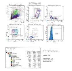
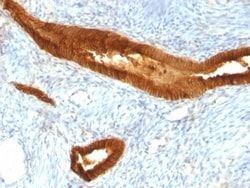
Flow Cytometry, Immunohistochemistry (Frozen), Immunofluorescence
Conjugate
Unconjugated
Host Species
Mouse
Research Discipline
Adaptive Immunity, Asthma, Immunology
Formulation
10mM PBS and 0.05% BSA with 0.05% Sodium Azide
Gene ID (Entrez)
3117
Immunogen
T4-positive CTL clone HG-38
Primary or Secondary
Primary
Content And Storage
Store at 4C.
Molecular Weight of Antigen
60 kDa
Clone
SPV-L3
Dilution
Flow Cytometry 0.5 - 1 ug/million cells in 0.1 ml, Immunohistochemistry-Frozen 0.5 - 1.0 ug/ml, Immunofluorescence 0.5 - 1.0 ug/ml
Classification
Monoclonal
Form
Purified
Regulatory Status
RUO
Target Species
Human, Porcine
Gene Alias
CD, CELIAC1DQ alpha 1 chain, DC-1 alpha chain, DQ-A1, FLJ27088, FLJ27328, GSE, HLA class II histocompatibility antigen, DQ(W3) alpha chain, HLA-DCA, HLA-DQA, leucocyte antigen DQA1, leukocyte antigen alpha chain, major histocompatibility complex, class II, DQ alpha 1, MGC149527, MHC class II antigen, MHC class II DQA1, MHC class II HLA-D alpha glycoprotein, MHC class II HLA-DQ-alpha-1, MHC class II surface glycoprotein, MHC HLA-DQ alpha
Gene Symbols
HLA-DQ
Isotype
IgG2a κ
Purification Method
Protein A or G purified
Test Specificity
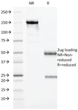
Recognizes a DQ antigen, which is a dimer of 60kDa. The class II molecule is a heterodimer consisting of an alpha (DQA) and a beta chain (DQB), both anchored in the membrane. It plays a central role in the immune system by presenting peptides derived from extracellular proteins. Class II molecules are expressed in antigen presenting cells (APC: B Lymphocytes, dendritic cells, macrophages). The alpha chain is approximately 33-35kDa. It is encoded by 5 exons; exon 1 encodes the leader peptide, exons 2 and 3 encode the two extracellular domains, and exon 4 encodes the transmembrane domain and the cytoplasmic tail. Within the DQ molecule both the alpha chain and the beta chain contain the polymorphisms specifying the peptide binding specificities, resulting in up to four different molecules. Typing for these polymorphisms is routinely done for bone marrow transplantation. This MAb strongly blocks cytotoxicity activity of T4-positive cytotoxic T cell clones.
Related Products


Description
- Ensure accurate, reproducible results in Flow Cytometry, Immunohistochemistry (Paraffin), Immunofluorescence HLA DQ Monoclonal specifically detects HLA DQ in Human, Porcine samples
- It is validated for Flow Cytometry, Immunocytochemistry/Immunofluorescence, Immunofluorescence.
Compare Similar Items
Show Difference
Antigen: HLA DQ
Concentration: 0.2 mg/mL
Applications: Flow Cytometry, Immunohistochemistry (Frozen), Immunofluorescence
Conjugate: Unconjugated
Host Species: Mouse
Research Discipline: Adaptive Immunity, Asthma, Immunology
Formulation: 10mM PBS and 0.05% BSA with 0.05% Sodium Azide
Gene ID (Entrez): 3117
Immunogen: T4-positive CTL clone HG-38
Primary or Secondary: Primary
Content And Storage: Store at 4C.
Molecular Weight of Antigen: 60 kDa
Clone: SPV-L3
Dilution: Flow Cytometry 0.5 - 1 ug/million cells in 0.1 ml, Immunohistochemistry-Frozen 0.5 - 1.0 ug/ml, Immunofluorescence 0.5 - 1.0 ug/ml
Classification: Monoclonal
Form: Purified
Regulatory Status: RUO
Target Species: Human, Porcine
Gene Alias: CD, CELIAC1DQ alpha 1 chain, DC-1 alpha chain, DQ-A1, FLJ27088, FLJ27328, GSE, HLA class II histocompatibility antigen, DQ(W3) alpha chain, HLA-DCA, HLA-DQA, leucocyte antigen DQA1, leukocyte antigen alpha chain, major histocompatibility complex, class II, DQ alpha 1, MGC149527, MHC class II antigen, MHC class II DQA1, MHC class II HLA-D alpha glycoprotein, MHC class II HLA-DQ-alpha-1, MHC class II surface glycoprotein, MHC HLA-DQ alpha
Gene Symbols: HLA-DQ
Isotype: IgG2a κ
Purification Method: Protein A or G purified
Test Specificity: Recognizes a DQ antigen, which is a dimer of 60kDa. The class II molecule is a heterodimer consisting of an alpha (DQA) and a beta chain (DQB), both anchored in the membrane. It plays a central role in the immune system by presenting peptides derived from extracellular proteins. Class II molecules are expressed in antigen presenting cells (APC: B Lymphocytes, dendritic cells, macrophages). The alpha chain is approximately 33-35kDa. It is encoded by 5 exons; exon 1 encodes the leader peptide, exons 2 and 3 encode the two extracellular domains, and exon 4 encodes the transmembrane domain and the cytoplasmic tail. Within the DQ molecule both the alpha chain and the beta chain contain the polymorphisms specifying the peptide binding specificities, resulting in up to four different molecules. Typing for these polymorphisms is routinely done for bone marrow transplantation. This MAb strongly blocks cytotoxicity activity of T4-positive cytotoxic T cell clones.
Antigen: EpCAM/TROP1
Concentration: 0.2mg/mL
Applications: Western Blot, Flow Cytometry, Immunohistochemistry (Paraffin), SDS-Page, Immunofluorescence
Conjugate: Unconjugated
Host Species: Mouse
Research Discipline: Cancer, Cancer Stem Cells
Formulation: 10mM PBS and 0.05% BSA with 0.05% Sodium Azide
Gene ID (Entrez): 4072
Immunogen: Neuraminidase treated GLS-1 human small cell lung carcinoma cells
Primary or Secondary: Primary
Content And Storage: Store at 4C.
Molecular Weight of Antigen: 41 kDa
Clone: MOC-31
Dilution: Western Blot 1:100 - 1:200, Flow Cytometry 5 - 10 ul/million cells in 0.1ml, Immunohistochemistry-Paraffin 1:100 - 1:200, SDS-Page, Immunofluorescence 1:50 - 1:100
Classification: Monoclonal
Form: Purified
Regulatory Status: RUO
Target Species: Human, Rat (Negative)
Gene Alias: 17-1A, 323/A3, ACSTD1, antigen identified by monoclonal AUA1, CD326 antigen, Cell surface glycoprotein Trop-1, chromosome 4, surface marker (35kD glycoprotein), DIAR5, EGP, EGP-2, EGP314, EGP40, EpCAM, epithelial cell adhesion molecule, Epithelial cell surface antigen, Epithelial glycoprotein, Epithelial glycoprotein 314, ESA, GA733-2EGP34, hEGP314, HNPCC8, KS 1/4 antigen, KS1/4, KSAHEA125, M1S2, M4S1Ly74, Major gastrointestinal tumor-associated protein GA733-2, MIC18MH99, MOC31, TACST-1, TACSTD1, TROP1CD326, Tumor-associated calcium signal transducer 1CO-17A
Gene Symbols: EPCAM
Isotype: IgG1 κ
Purification Method: Protein A or G purified
Test Specificity: Binding epitope of this antibody is located in the first EGF-like repeat domain (EGF1) between amino acids 27-59 of Ep-CAM. EGP40 is a 40-43kDa transmembrane epithelial glycoprotein, also identified as epithelial specific antigen (ESA), or epithelial cellular adhesion molecule (Ep-CAM). It is expressed on baso-lateral cell surface in most simple epithelia and a vast majority of carcinomas with the exception of adult squamous epithelium, hepatocytes and gastric epithelial cells. This antibody has been used to distinguish adenocarcinoma from pleural mesothelioma and hepatocellular carcinoma. This antibody is also useful in distinguishing serous carcinomas of the ovary from mesothelioma.
Antigen: __
Concentration: __
Applications: __
Conjugate: __
Host Species: __
Research Discipline: __
Formulation: __
Gene ID (Entrez): __
Immunogen: __
Primary or Secondary: __
Content And Storage: __
Molecular Weight of Antigen: __
Clone: __
Dilution: __
Classification: __
Form: __
Regulatory Status: __
Target Species: __
Gene Alias: __
Gene Symbols: __
Isotype: __
Purification Method: __
Test Specificity: __