Ki-67 Monoclonal Antibody (SolA15), eFluor™ 570, eBioscience™, Invitrogen™
Rat Monoclonal Antibody
Manufacturer: Fischer Scientific
The price for this product is unavailable. Please request a quote
Antigen
Ki-67
Concentration
0.2 mg/mL
Classification
Monoclonal
Form
Liquid
Regulatory Status
RUO
Formulation
PBS with 0.09% sodium azide; pH 7.2
Gene Alias
antigen identified by monoclonal antibody Ki 67; antigen identified by monoclonal antibody Ki-67; Antigen identified by monoclonal antibody Ki-67 homolog; Antigen KI-67; Antigen KI-67 homolog; antigen KI-67; proliferation marker protein Ki-67; antigen KI-67-like; cb31; D630048A14Rik; I79_022666; Ki67; Ki-67; KIA; LOW QUALITY PROTEIN: proliferation marker protein Ki-67; marker of proliferation Ki-67; MIB-; MIB-1; Mki67; PPP1R105; Proliferation marker protein Ki-67; proliferation-related Ki-67 antigen; protein phosphatase 1, regulatory subunit 105; RP11-380J17.2; sb:cb31; si:ch211-250b22.7; unnamed protein product; wu:fa11g09; wu:fb57a07; wu:fi14e05
Gene Symbols
Mki67
Primary or Secondary
Primary
Content And Storage
4° C, store in dark, DO NOT FREEZE!
Gene
Mki67
Clone
SolA15
Applications
Immunocytochemistry, Immunohistochemistry (Frozen), Immunohistochemistry (Paraffin)
Conjugate
eFluor 570
Host Species
Rat
Target Species
Canine, Cynomolgus Monkey, Human, Mouse, Non-human Primate, Rat
Gene Accession No.
E9PVX6, P46013
Gene ID (Entrez)
100686578, 102135895, 17345, 291234, 4288
Isotype
IgG2a κ
Purification Method
Affinity chromatography
Product Type
Antibody
Description
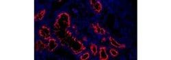
- Description: The monoclonal antibody SolA15 recognizes mouse and rat Ki-67, a 300 kDa nuclear protein
- Ki-67 is present during all active phases of the cell cycle (G1, S, G2, and mitosis), but is absent from resting cells (G0)
- Ki-67 is detected within the nucleus during interphase but redistributes to the chromosomes during mitosis
- Ki-67 is used as a marker for determining the growth fraction of a given population of cells
- In studies of tumor cells, the Ki-67 labeling index refers to the number of Ki-67 positive cells within the population and this is used to predict outcome of particular cancer types
- Ki-67 has been shown to interact with the DNA-bound protein chromobox protein homolog 3 (CBX3) (heterochromatin)
- The SolA15 antibody also recognizes human, non-human primate and canine Ki-67
- Applications Reported: This SolA15 antibody has been reported for use in immunohistochemical staining of frozen tissue sections, immunohistochemical staining of formalin-fixed paraffin embedded tissue sections, and immunocytochemistry
- Applications Tested: This SolA15 antibody has been tested immunocytochemistry on fixed and permeabilized C2C12 cells and can be used at less than or equal to 1 μg/mL
- It is recommended that the antibody be carefully titrated for optimal performance in the assay of interest
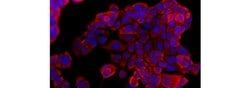
- Ki-67 is a nuclear protein that is expressed during various stages in the cell cycle, particularly during late G1, S, G2, and M phases
- The protein has a forkhead associated domain (FHA) through which it associates with euchromatin at the perichromosomal layer, the centromeric heterochromatin, and the nucleolus
- Ki-67 is shown to have a cell cycle dependent topographical distribution with perinucleolar expression at G1, expression in the nuclear matrix at G2, and expression on the chromosomes during M phase
- Ki-67 is commonly used as a proliferation marker because it is not detected in G0 cells, but increases steadily from G1 through mitosis
- Ki-67 antibodies are useful in establishing the cell growing fraction in neoplasms
- In neoplastic tissues, the prognostic value is comparable to the tritiated thymidine-labelling index
- The correlation between low Ki-67 index and histologically low-grade tumors is strong
- Ki-67 is routinely used as a neuronal marker of cell cycling and proliferation.
Compare Similar Items
Show Difference

Antigen: Ki-67
Concentration: 0.2 mg/mL
Classification: Monoclonal
Form: Liquid
Regulatory Status: RUO
Formulation: PBS with 0.09% sodium azide; pH 7.2
Gene Alias: antigen identified by monoclonal antibody Ki 67; antigen identified by monoclonal antibody Ki-67; Antigen identified by monoclonal antibody Ki-67 homolog; Antigen KI-67; Antigen KI-67 homolog; antigen KI-67; proliferation marker protein Ki-67; antigen KI-67-like; cb31; D630048A14Rik; I79_022666; Ki67; Ki-67; KIA; LOW QUALITY PROTEIN: proliferation marker protein Ki-67; marker of proliferation Ki-67; MIB-; MIB-1; Mki67; PPP1R105; Proliferation marker protein Ki-67; proliferation-related Ki-67 antigen; protein phosphatase 1, regulatory subunit 105; RP11-380J17.2; sb:cb31; si:ch211-250b22.7; unnamed protein product; wu:fa11g09; wu:fb57a07; wu:fi14e05
Gene Symbols: Mki67
Primary or Secondary: Primary
Content And Storage: 4° C, store in dark, DO NOT FREEZE!
Gene: Mki67
Clone: SolA15
Applications: Immunocytochemistry, Immunohistochemistry (Frozen), Immunohistochemistry (Paraffin)
Conjugate: eFluor 570
Host Species: Rat
Target Species: Canine, Cynomolgus Monkey, Human, Mouse, Non-human Primate, Rat
Gene Accession No.: E9PVX6, P46013
Gene ID (Entrez): 100686578, 102135895, 17345, 291234, 4288
Isotype: IgG2a κ
Purification Method: Affinity chromatography
Product Type: Antibody
Antigen: Pan Cytokeratin
Concentration: 0.2 mg/mL
Classification: Monoclonal
Form: Liquid
Regulatory Status: RUO
Formulation: PBS with 0.09% sodium azide; pH 7.2
Gene Alias: 2310016L08Rik; 3300001P10Rik; 39.1; 40-kDa keratin intermediate filament; 47 kDa cytokeratin; 56 kDa cytokeratin; 57kd keratin; 57kDa keratin; 58 kDa cytokeratin; 60-kDa keratin; 63kDa Keratin; 65 kDa cytokeratin; 67 kDa cytokeratin; AA960620; adult keratin; AI324768; AI528832; AI626930; AI663979; AL022697; alpha keratin; AU019895; AW108092; AW146334; basic epidermal type II cytokeratin (carboxy-terminal region, clone pUF164); BB005427; BCIE; BIE; Card2; cell proliferation-inducing gene 46 protein; CK 2e; CK1; CK-1; CK10; CK-10; CK13; CK-13; CK14; CK-14; CK15; CK-15; CK16; CK-16; CK-17; CK18; CK-18; CK19; CK-19; CK-1B; CK-2e; CK3; CK-3; CK4; CK-4; CK5; CK-5; ck55; CK6A; CK-6A; CK6C; CK-6C; CK6D; CK-6D; CK-6E; CK7; CK-7; CK8; CK-8; CYK18; CYK4; CYK8; CYKER; cytokeratin 1; cytokeratin 10; cytokeratin 13; cytokeratin 14; cytokeratin 15; cytokeratin 16; cytokeratin 18; cytokeratin 19; cytokeratin 3; cytokeratin 4; cytokeratin 5; cytokeratin 6A; cytokeratin 6C; cytokeratin 6D; cytokeratin 7; cytokeratin 8; cytokeratin 8 (370 AA); cytokeratin endo A; Cytokeratin endo B; cytokeratin otokeratin; cytokeratin type II; cytokeratin type II, component Ib/c; cytokeratin type II, component III; cytokeratin VIB; cytokeratin VII; cytokeratin-1; Cytokeratin-10; cytokeratin-13; Cytokeratin-14; cytokeratin-15; Cytokeratin-16; cytokeratin-17; Cytokeratin-18; cytokeratin-19; Cytokeratin-1B; Cytokeratin-2e; cytokeratin-3; Cytokeratin-4; cytokeratin-5; Cytokeratin-6A; cytokeratin-6B; cytokeratin-6C; Cytokeratin-6D; cytokeratin-6E; cytokeratin-7; Cytokeratin-8; cytokeratin-A; Cytoskeletal 57 kDa keratin; D130054E02Rik; D15Wsu77e; DDD; DDD1; ear specific cytokeratin; EBS2; ebs3; ebs4; EGK_03684; EGK_03685; EHK; EHK1; Endo B; EndoA; EndoC; epidermal keratin 10; epidermal keratin VII; epidermolysis bullosa simplex 2 Dowling-Meara/Kobner/Weber-Cockayne types; epidermolysis bullosa simplex, Dowling-Meara, Koebner; epidermolytic hyperkeratosis 1; epidermolytic hyperkeratosis; keratosis palmaris et plantaris; epithelial keratin 1; epithelial keratin 10; epithelial keratin 2e; Epithelial keratin-1; Epithelial keratin-2e; EPPK; fgk; fin and gill keratin; FNEPPK; focal non-epidermolytic palmoplantar keratoderma; GK-19; Hair alpha protein; HMWCK; Hom s 5; I79_019823; I79_021074; I79_023185; I79_024335; intermediate filament protein; K1; K10; K13; K14; K15; K16; K17; K18; K19; K1B; K1C1; K1CO; K1CP; K1CS; K2C7; K2C8; K2e; K3; K3 keratin; K4; K5; K6A; K6a keratin; K6C; K6D; K7; K77; K8; Ka10; Ka13; Ka14; Ka15; Ka16; Ka17; Ka19; kamp-keratin derived antimicrobial peptide; Kb1; Kb2; Kb4; Kb7; KDAMP; KER1; Ker10; Ker2; KERA; keratin; keratin 1; keratin 1 (epidermolytic hyperkeratosis); keratin 1, type II; keratin 10; keratin 10 (epidermolytic hyperkeratosis); keratin 10 (epidermolytic hyperkeratosis; keratosis palmaris et plantaris); keratin 10, type I; keratin 10, type I L homeolog; keratin 10, type I S homeolog; keratin 12, gene 2 S homeolog; keratin 13; keratin 13, type I; keratin 13, type I S homeolog; keratin 14; keratin 14 (epidermolysis bullosa simplex, Dowling-Meara, Koebner); keratin 14, type I; keratin 14, type I L homeolog; keratin 15; keratin 15, gene 1 S homeolog; keratin 15, type I; keratin 16; keratin 16 (focal non-epidermolytic palmoplantar keratoderma); keratin 16, type I; keratin 16, type I S homeolog; keratin 17; keratin 17 L homeolog; keratin 17, type I; keratin 17, type I L homeolog; keratin 18; keratin 18, type I; keratin 19; keratin 19 S homeolog; keratin 19 S homeolog; keratin 19; keratin 19, type I; Keratin 1B; keratin 2; keratin 2 (epidermal ichthyosis bullosa of Siemens); keratin 2 epidermis; keratin 2, type II; keratin 24; keratin 2A; keratin 2A (epidermal ichthyosis bullosa of Siemens); keratin 3; keratin 3, type II; keratin 4; keratin 4, type II; keratin 5; keratin 5 (epidermolysis bullosa simplex Dowling-Meara/Kobner/Weber-Cockayne types); keratin 5 (epidermolysis bullosa simplex, Dowling-Meara/Kobner/Weber-Cockayne types)
Gene Symbols: KRT1, KRT10, KRT13, KRT14, KRT15, KRT16, KRT17, KRT18, KRT19, KRT2, KRT3, KRT4, KRT5, Krt6a, KRT7, KRT76, KRT77, KRT8
Primary or Secondary: Primary
Content And Storage: 4° C, store in dark, DO NOT FREEZE!
Gene: __
Clone: AE1/AE3
Applications: Immunocytochemistry, Immunohistochemistry (Frozen), Immunohistochemistry (Paraffin)
Conjugate: eFluor 570
Host Species: Mouse
Target Species: Canine, Human, Mouse, Non-human Primate, Rabbit, Rhesus Monkey
Gene Accession No.: P02533, P02535, P02538, P04104, P04264, P05783, P05784, P05787, P07744, P08727, P08729, P08730, P08779, P11679, P12035, P13645, P13646, P13647, P19001, P19012, P19013, P35908, P50446, Q01546, Q04695, Q29426, Q3TTY5, Q3UV17, Q61414, Q61781, Q6EIY9, Q6EIZ0, Q6EIZ1, Q6IFZ6, Q7Z794, Q922U2, Q9DCV7, Q9QWL7, Q9Z2K1
Gene ID (Entrez): 100009029, 100341120, 100341376, 100344183, 100344942, 100345579, 100348388, 100348891, 100352658, 100353162, 100353415, 100353668, 100354174, 100424400, 100425096, 100426180, 100426330, 100682832, 100683333, 100683401, 100685323, 100685404, 100685481, 100687760, 103348273, 106994041, 110308, 110310, 16661, 16663, 16664, 16665, 16666, 16667, 16668, 16669, 16678, 16681, 16682, 16687, 16691, 374454, 3848, 3849, 3850, 3851, 3852, 3853, 3855, 3856, 3858, 3860, 3861, 3866, 3868, 3872, 3875, 3880, 406220, 444850, 444857, 477601, 477602, 486513, 486518, 486523, 490977, 490978, 490983, 491006, 51350, 697425, 699194, 699320, 699567, 700060, 700449, 702153, 705390, 706830, 707236, 718983, 77055
Isotype: IgG1
Purification Method: Affinity chromatography
Product Type: Antibody
Antigen: CD4
Concentration: 0.2 mg/mL
Classification: Monoclonal
Form: Liquid
Regulatory Status: RUO
Formulation: PBS with 0.09% sodium azide; pH 7.2
Gene Alias: Activation B7-1 antigen; B7; B7.1; B7-1; BB1; B-lymphocyte activation antigen B7; CD28LG; CD28LG1; CD4; CD4 antigen; CD4 antigen (p55); CD4 antigen p55; Cd4 molecule; CD4 precursor; CD4 receptor; CD4, allele 1; cd4a; CD4mut; CD80; CD80 antigen (CD28 antigen ligand 1, B7-1 antigen); CD80 molecule; cell surface glycoprotein CD4; costimulatory factor CD80; costimulatory molecule variant IgV-CD80; CTLA-4 counter-receptor B7.1; fCD4; L3T4; LAB7; Leu-3; Ly-4; lymphocyte antigen CD4; lymphocyte antigen CD4 precursor; membrane protein; p55; T-cell differentiation antigen L3T4; T-cell surface antigen T4/Leu-3; T-cell surface glycoprotein CD4; T-cell surface glycoprotein CD4 precursor (T-cell surface antigen T4/Leu-3) (T-cell differentiation antigen L3T4); T-lymphocyte activation antigen CD80; W3/25; W3/25 antigen
Gene Symbols: CD4
Primary or Secondary: Primary
Content And Storage: 4° C, store in dark, DO NOT FREEZE!
Gene: CD4
Clone: RM4-5
Applications: Immunocytochemistry, Immunohistochemistry (Frozen)
Conjugate: eFluor 615
Host Species: Rat
Target Species: Mouse
Gene Accession No.: P06332
Gene ID (Entrez): 12504
Isotype: IgG2a κ
Purification Method: Affinity chromatography
Product Type: Antibody