ErbB2/Her2 Antibody (HRB2/718), Novus Biologicals™
Mouse Monoclonal Antibody
Manufacturer: Fischer Scientific
The price for this product is unavailable. Please request a quote
Antigen
ErbB2/Her2
Dilution
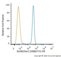
Flow Cytometry 0.5-1ug/million cells, Immunocytochemistry/Immunofluorescence 0.5-1ug/ml
Classification
Monoclonal
Form
Purified
Regulatory Status
RUO
Target Species
Human
Gene Accession No.
P04626
Gene ID (Entrez)
2064
Immunogen
Recombinant extracellular domain of human HER-2 protein
Primary or Secondary
Primary
Content And Storage
Store at 4C.
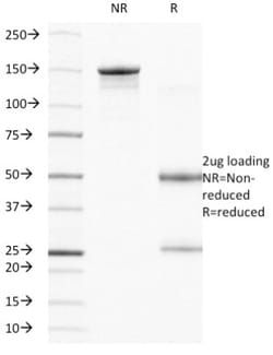
Molecular Weight of Antigen
185 kDa
Clone
HRB2/718
Applications
Flow Cytometry, Immunocytochemistry, Immunofluorescence
Conjugate
Unconjugated
Host Species
Mouse
Research Discipline
Breast Cancer, Cancer, Cellular Markers, Core ESC Like Genes, Phospho Specific, Protein Kinase, Stem Cell Markers, Tumor Suppressors
Formulation
PBS with 0.05% BSA. with 0.05% Sodium Azide
Gene Alias
CD340, CD340 antigen, c-erb B2/neu protein, EC 2.7.10, EGFR2, HER-2, HER2EC 2.7.10.1, herstatin, Metastatic lymph node gene 19 protein, MLN 19, MLN19, NEUHER-2/neu, neuroblastoma/glioblastoma derived oncogene homolog, NGLTKR1, p185erbB2, Proto-oncogene c-ErbB-2, Proto-oncogene Neu, receptor tyrosine-protein kinase erbB-2, Tyrosine kinase-type cell surface receptor HER2, v-erb-b2 avian erythroblastic leukemia viral oncogene homolog 2(neuro/glioblastoma derived oncogene homolog), v-erb-b2 erythroblastic leukemia viral oncogene homolog 2, neuro/glioblastomaderived oncogene homolog (avian)
Gene Symbols
ERBB2
Isotype
IgG1 κ
Purification Method
Protein A purified
Test Specificity
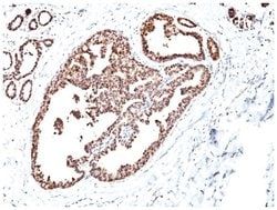
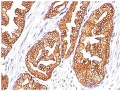
Recognizes a protein of 185kDa, which is identified as c-erbB-2/HER-2/neu. Its epitope is localized in the extracellular domain. C-erbB-2/HER-2 is a member of the EGFR family. This MAb is specific and shows minimal cross-reaction with other members of the EGFR-family. Receptors of this family are located on the plasma membrane and consist of an extracellular ligand-binding domain that is connected to a large intracellular domain by a single transmembrane sequence. c-erbB-2/HER-2 protein is over-expressed in a variety of carcinomas especially those of breast and ovary.
Related Products


Description
- ErbB2/Her2 Monoclonal specifically detects ErbB2/Her2 in Human samples
- It is validated for Flow Cytometry, ELISA, Immunocytochemistry/Immunofluorescence, Protein Array, Flow (Intracellular).
Compare Similar Items
Show Difference
Antigen: ErbB2/Her2
Dilution: Flow Cytometry 0.5-1ug/million cells, Immunocytochemistry/Immunofluorescence 0.5-1ug/ml
Classification: Monoclonal
Form: Purified
Regulatory Status: RUO
Target Species: Human
Gene Accession No.: P04626
Gene ID (Entrez): 2064
Immunogen: Recombinant extracellular domain of human HER-2 protein
Primary or Secondary: Primary
Content And Storage: Store at 4C.
Molecular Weight of Antigen: 185 kDa
Clone: HRB2/718
Applications: Flow Cytometry, Immunocytochemistry, Immunofluorescence
Conjugate: Unconjugated
Host Species: Mouse
Research Discipline: Breast Cancer, Cancer, Cellular Markers, Core ESC Like Genes, Phospho Specific, Protein Kinase, Stem Cell Markers, Tumor Suppressors
Formulation: PBS with 0.05% BSA. with 0.05% Sodium Azide
Gene Alias: CD340, CD340 antigen, c-erb B2/neu protein, EC 2.7.10, EGFR2, HER-2, HER2EC 2.7.10.1, herstatin, Metastatic lymph node gene 19 protein, MLN 19, MLN19, NEUHER-2/neu, neuroblastoma/glioblastoma derived oncogene homolog, NGLTKR1, p185erbB2, Proto-oncogene c-ErbB-2, Proto-oncogene Neu, receptor tyrosine-protein kinase erbB-2, Tyrosine kinase-type cell surface receptor HER2, v-erb-b2 avian erythroblastic leukemia viral oncogene homolog 2(neuro/glioblastoma derived oncogene homolog), v-erb-b2 erythroblastic leukemia viral oncogene homolog 2, neuro/glioblastomaderived oncogene homolog (avian)
Gene Symbols: ERBB2
Isotype: IgG1 κ
Purification Method: Protein A purified
Test Specificity: Recognizes a protein of 185kDa, which is identified as c-erbB-2/HER-2/neu. Its epitope is localized in the extracellular domain. C-erbB-2/HER-2 is a member of the EGFR family. This MAb is specific and shows minimal cross-reaction with other members of the EGFR-family. Receptors of this family are located on the plasma membrane and consist of an extracellular ligand-binding domain that is connected to a large intracellular domain by a single transmembrane sequence. c-erbB-2/HER-2 protein is over-expressed in a variety of carcinomas especially those of breast and ovary.
Antigen: HSP27
Dilution: Western Blot 0.25-0.5ug/ml, Flow Cytometry 0.5-1ug/million cells, Immunocytochemistry/Immunofluorescence 0.5-1ug/ml, Immunohistochemistry-Paraffin 0.5-1.0ug/ml
Classification: Monoclonal
Form: Purified
Regulatory Status: RUO
Target Species: Human, Mouse, Rat, Chicken, Primate, Sheep
Gene Accession No.: P04792
Gene ID (Entrez): 3315
Immunogen: Partially purified hsp27 (earlier called 24K) protein from breast cancer MCF-7 cells.
Primary or Secondary: Primary
Content And Storage: Store at 4C.
Molecular Weight of Antigen: 27 kDa
Clone: SPM252
Applications: Western Blot, Flow Cytometry, Immunocytochemistry, Immunofluorescence, Immunohistochemistry (Paraffin)
Conjugate: Unconjugated
Host Species: Mouse
Research Discipline: Cancer, Cell Biology, Cell Cycle and Replication, Golgi Apparatus Markers, Membrane Trafficking and Chaperones, Neuroscience, Phospho Specific
Formulation: PBS with 0.05% BSA. with 0.05% Sodium Azide
Gene Alias: 28 kDa heat shock protein, DKFZp586P1322, Estrogen-regulated 24 kDa protein, Heat shock 27 kDa protein, heat shock 27kD protein 1, heat shock 27kDa protein 1, heat shock protein beta-1, HMN2B, HS.76067, Hsp25, HSP27HSP 27, HSP28CMT2F, HspB1, SRP27, Stress-responsive protein 27
Gene Symbols: HSPB1
Isotype: IgG1 κ
Purification Method: Protein A purified
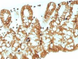
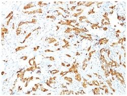
Test Specificity: It recognizes a 24-27kDa estrogen-regulated protein, identified as heat shock protein 27 (hsp27). Hsp27 was recently found to be identical to the estrogen-induced p29 and 24K protein. About 50% of breast carcinomas are positive for hsp27 especially those that are also positive for estrogen and/or progesterone receptor. HSP27 has also been implicated in drug resistance in cancer cells.
Antigen: HSP27
Dilution: Western Blot 0.25-0.5ug/ml, Flow Cytometry 0.5-1ug/million cells, Immunocytochemistry/Immunofluorescence 0.5-1ug/ml, Immunohistochemistry-Paraffin 0.5-1.0ug/ml
Classification: Monoclonal
Form: Purified
Regulatory Status: RUO
Target Species: Human, Mouse, Rat, Chicken, Primate, Sheep
Gene Accession No.: P04792
Gene ID (Entrez): 3315
Immunogen: Partially purified hsp27 (earlier called 24K) protein from breast cancer MCF-7 cells.
Primary or Secondary: Primary
Content And Storage: Store at 4C.
Molecular Weight of Antigen: 27 kDa
Clone: SPM252
Applications: Western Blot, Flow Cytometry, Immunocytochemistry, Immunofluorescence, Immunohistochemistry (Paraffin)
Conjugate: Unconjugated
Host Species: Mouse
Research Discipline: Cancer, Cell Biology, Cell Cycle and Replication, Golgi Apparatus Markers, Membrane Trafficking and Chaperones, Neuroscience, Phospho Specific
Formulation: PBS with 0.05% BSA. with 0.05% Sodium Azide
Gene Alias: 28 kDa heat shock protein, DKFZp586P1322, Estrogen-regulated 24 kDa protein, Heat shock 27 kDa protein, heat shock 27kD protein 1, heat shock 27kDa protein 1, heat shock protein beta-1, HMN2B, HS.76067, Hsp25, HSP27HSP 27, HSP28CMT2F, HspB1, SRP27, Stress-responsive protein 27
Gene Symbols: HSPB1
Isotype: IgG1 κ
Purification Method: Protein A purified
Test Specificity: It recognizes a 24-27kDa estrogen-regulated protein, identified as heat shock protein 27 (hsp27). Hsp27 was recently found to be identical to the estrogen-induced p29 and 24K protein. About 50% of breast carcinomas are positive for hsp27 especially those that are also positive for estrogen and/or progesterone receptor. HSP27 has also been implicated in drug resistance in cancer cells.