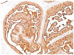
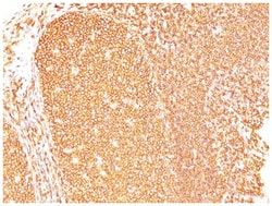
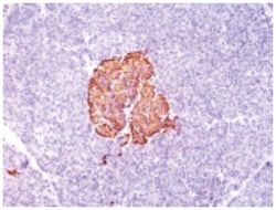
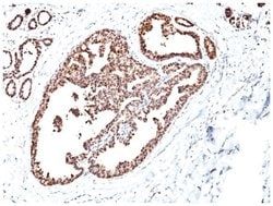
HSP60 Mouse, Clone: SPM253, Novus Biologicals™
Mouse Monoclonal Antibody
Manufacturer: Fischer Scientific
The price for this product is unavailable. Please request a quote
Antigen
HSP60
Dilution
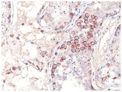
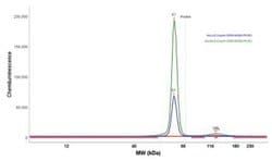
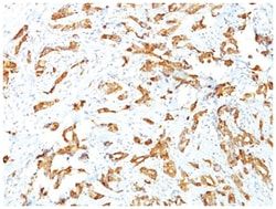
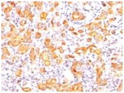
Western Blot 0.25-0.5ug/ml, Flow Cytometry 0.5-1ug/million cells, Immunocytochemistry/Immunofluorescence 0.5-1ug/ml, Immunohistochemistry-Paraffin 0.5-1.0ug/ml
Classification
Monoclonal
Form
Purified
Regulatory Status
RUO
Target Species
Human, Mouse, Rat, Porcine, Bovine, Canine, Chicken, Drosophila, Hamster, Primate, Rabbit, Sheep, Xenopus species
Gene Accession No.
P10809
Gene ID (Entrez)
3329
Immunogen
Recombinant human HSP60 protein
Primary or Secondary
Primary
Content And Storage
Store at 4C.
Molecular Weight of Antigen
60 kDa
Clone
SPM253
Applications
Western Blot, Flow Cytometry, Immunocytochemistry, Immunofluorescence, Immunohistochemistry (Paraffin)
Conjugate
Unconjugated
Host Species
Mouse
Research Discipline
Apoptosis, Cellular Markers, Membrane Trafficking and Chaperones, Mitochondrial Markers
Formulation
PBS with 0.05% BSA. with 0.05% Sodium Azide
Gene Alias
60 kDa chaperonin, Chaperonin 60, CPN60, GROEL, heat shock 60kD protein 1 (chaperonin), heat shock 60kDa protein 1 (chaperonin), Heat shock protein 60, heat shock protein 65, HLD4, Hsp60, HSP-60, HSP60SPG13, HSP65, HuCHA60, Mitochondrial matrix protein P1,60 kDa heat shock protein, mitochondrial, P60 lymphocyte protein, short heat shock protein 60 Hsp60s1, spastic paraplegia 13 (autosomal dominant)
Gene Symbols
HSPD1
Isotype
IgG1 κ
Purification Method
Protein A purified
Test Specificity
Recognizes a 60kDa protein, identified as the heat shock protein 60 (hsp60). Its epitope is localized between aa 383-447 of human hsp60. A wide variety of environmental and pathophysiological stressful conditions trigger the synthesis of a family of proteins known as heat shock proteins (hsps), more appropriately called as stress response proteins (srps). hsp60 is a potential antigen in a number of autoimmune diseases. In human arthritis and in experimentally induced arthritis in animals, disease development coincides with the development of immune reactivity directed against not only bacterial hsp60, but also against its mammalian homolog.
Related Products

Description
- HSP60 Monoclonal specifically detects HSP60 in Human, Mouse, Rat, Porcine, Bovine, Canine, Chicken, Drosophila, Hamster, Monkey, Rabbit, Sheep, Xenopus samples
- It is validated for Western Blot, Flow Cytometry, Immunohistochemistry, Immunocytochemistry/Immunofluorescence, Immunohistochemistry-Paraffin.
Compare Similar Items
Show Difference
Antigen: HSP60
Dilution: Western Blot 0.25-0.5ug/ml, Flow Cytometry 0.5-1ug/million cells, Immunocytochemistry/Immunofluorescence 0.5-1ug/ml, Immunohistochemistry-Paraffin 0.5-1.0ug/ml
Classification: Monoclonal
Form: Purified
Regulatory Status: RUO
Target Species: Human, Mouse, Rat, Porcine, Bovine, Canine, Chicken, Drosophila, Hamster, Primate, Rabbit, Sheep, Xenopus species
Gene Accession No.: P10809
Gene ID (Entrez): 3329
Immunogen: Recombinant human HSP60 protein
Primary or Secondary: Primary
Content And Storage: Store at 4C.
Molecular Weight of Antigen: 60 kDa
Clone: SPM253
Applications: Western Blot, Flow Cytometry, Immunocytochemistry, Immunofluorescence, Immunohistochemistry (Paraffin)
Conjugate: Unconjugated
Host Species: Mouse
Research Discipline: Apoptosis, Cellular Markers, Membrane Trafficking and Chaperones, Mitochondrial Markers
Formulation: PBS with 0.05% BSA. with 0.05% Sodium Azide
Gene Alias: 60 kDa chaperonin, Chaperonin 60, CPN60, GROEL, heat shock 60kD protein 1 (chaperonin), heat shock 60kDa protein 1 (chaperonin), Heat shock protein 60, heat shock protein 65, HLD4, Hsp60, HSP-60, HSP60SPG13, HSP65, HuCHA60, Mitochondrial matrix protein P1,60 kDa heat shock protein, mitochondrial, P60 lymphocyte protein, short heat shock protein 60 Hsp60s1, spastic paraplegia 13 (autosomal dominant)
Gene Symbols: HSPD1
Isotype: IgG1 κ
Purification Method: Protein A purified
Test Specificity: Recognizes a 60kDa protein, identified as the heat shock protein 60 (hsp60). Its epitope is localized between aa 383-447 of human hsp60. A wide variety of environmental and pathophysiological stressful conditions trigger the synthesis of a family of proteins known as heat shock proteins (hsps), more appropriately called as stress response proteins (srps). hsp60 is a potential antigen in a number of autoimmune diseases. In human arthritis and in experimentally induced arthritis in animals, disease development coincides with the development of immune reactivity directed against not only bacterial hsp60, but also against its mammalian homolog.
Antigen: HSP60
Dilution: Western Blot 0.25-0.5ug/ml, Simple Western 1:10, Flow Cytometry 0.5-1ug/million cells, Immunocytochemistry/Immunofluorescence 0.5-1ug/ml
Classification: Monoclonal
Form: Purified
Regulatory Status: RUO
Target Species: Human, Mouse, Rat, Bacteria, Chicken, Fungi, Guinea Pig, Hamster, Parasite, Primate
Gene Accession No.: P10809
Gene ID (Entrez): 3329
Immunogen: Recombinant human HSP60 protein
Primary or Secondary: Primary
Content And Storage: Store at 4C.
Molecular Weight of Antigen: 60 kDa
Clone: LK2
Applications: Western Blot, Flow Cytometry, Immunocytochemistry, Immunofluorescence
Conjugate: Unconjugated
Host Species: Mouse
Research Discipline: Apoptosis, Cellular Markers, Membrane Trafficking and Chaperones, Mitochondrial Markers
Formulation: PBS with 0.05% BSA. with 0.05% Sodium Azide
Gene Alias: 60 kDa chaperonin, Chaperonin 60, CPN60, GROEL, heat shock 60kD protein 1 (chaperonin), heat shock 60kDa protein 1 (chaperonin), Heat shock protein 60, heat shock protein 65, HLD4, Hsp60, HSP-60, HSP60SPG13, HSP65, HuCHA60, Mitochondrial matrix protein P1,60 kDa heat shock protein, mitochondrial, P60 lymphocyte protein, short heat shock protein 60 Hsp60s1, spastic paraplegia 13 (autosomal dominant)
Gene Symbols: HSPD1
Isotype: IgG1 κ
Purification Method: Protein A purified
Test Specificity: Recognizes a 60kDa protein, identified as the heat shock protein 60 (hsp60). Its epitope is localized between aa 383-419 of human hsp60. A wide variety of environmental and pathophysiological stressful conditions trigger the synthesis of a family of proteins known as heat shock proteins (hsps), more appropriately called as stress response proteins (srps). hsp60 is a potential antigen in a number of autoimmune diseases. In human arthritis and in experimentally induced arthritis in animals, disease development coincides with the development of immune reactivity directed against not only bacterial hsp60, but also against its mammalian homolog. Clone LK1, unlike LK2, recognizes only the mammalian (not bacterial) hsp60 and is useful in distinguishing hsp60 from mammals and bacteria.
Antigen: ECM-1/Secretory Component P85
Dilution: Flow Cytometry 0.5-1ug/million cells, Immunocytochemistry/Immunofluorescence 1-2ug/ml, Immunohistochemistry-Paraffin 0.5-1.0ug/ml
Classification: Monoclonal
Form: Purified
Regulatory Status: RUO
Target Species: Human, Rat
Gene Accession No.: Q16610
Gene ID (Entrez): 1893
Immunogen: Secretory Component protein isolated from human colostrum
Primary or Secondary: Primary
Content And Storage: Store at 4C.
Molecular Weight of Antigen: 80 kDa
Clone: SPM217
Applications: Flow Cytometry, Immunocytochemistry, Immunofluorescence, Immunohistochemistry (Paraffin)
Conjugate: Unconjugated
Host Species: Mouse
Research Discipline: Extracellular Matrix, Immunology
Formulation: PBS with 0.05% BSA. with 0.05% Sodium Azide
Gene Alias: ECM1, extracellular matrix protein 1, Secretory Component Glycoprotein, Secretory component p85
Gene Symbols: ECM1
Isotype: IgG1 κ
Purification Method: Protein A purified
Test Specificity: This MAb reacts with a reduction-resistant epitope present in both free and SIgA bound Secretory Component. It does not react with the cell lines lacking secretory component. The antibody is useful for studying the distribution and level of both free and bound secretory component. Secretory component is differentially expressed in epithelium, and the antibody is a popular marker for identifying subpopulations of epithelial cells and epithelial differentiation. The Secretory component antibody is a useful research tool for studying mucosal immunity, inflammation, remodeling, differentiation and tumorigenesis, all processes associated with differential secretory component expression.