ECM-1/Secretory Component P85 Mouse, Clone: SPM217, Novus Biologicals™
Mouse Monoclonal Antibody
Manufacturer: Fischer Scientific
The price for this product is unavailable. Please request a quote
Antigen
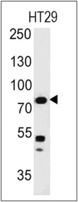
ECM-1/Secretory Component P85
Dilution
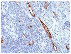
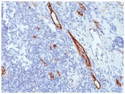
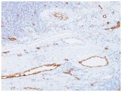
Flow Cytometry 0.5-1ug/million cells, Immunocytochemistry/Immunofluorescence 1-2ug/ml, Immunohistochemistry-Paraffin 0.5-1.0ug/ml
Classification
Monoclonal
Form
Purified
Regulatory Status
RUO
Target Species
Human, Rat
Gene Accession No.
Q16610
Gene ID (Entrez)
1893
Immunogen
Secretory Component protein isolated from human colostrum
Primary or Secondary
Primary
Content And Storage
Store at 4C.
Molecular Weight of Antigen
80 kDa
Clone
SPM217
Applications
Flow Cytometry, Immunocytochemistry, Immunofluorescence, Immunohistochemistry (Paraffin)
Conjugate
Unconjugated
Host Species
Mouse
Research Discipline
Extracellular Matrix, Immunology
Formulation
PBS with 0.05% BSA. with 0.05% Sodium Azide
Gene Alias
ECM1, extracellular matrix protein 1, Secretory Component Glycoprotein, Secretory component p85
Gene Symbols
ECM1
Isotype
IgG1 κ
Purification Method
Protein A purified
Test Specificity
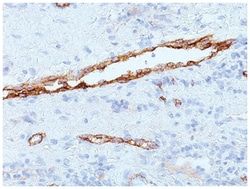
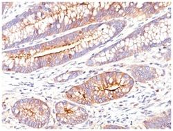
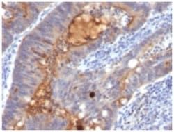
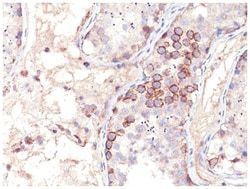
This MAb reacts with a reduction-resistant epitope present in both free and SIgA bound Secretory Component. It does not react with the cell lines lacking secretory component. The antibody is useful for studying the distribution and level of both free and bound secretory component. Secretory component is differentially expressed in epithelium, and the antibody is a popular marker for identifying subpopulations of epithelial cells and epithelial differentiation. The Secretory component antibody is a useful research tool for studying mucosal immunity, inflammation, remodeling, differentiation and tumorigenesis, all processes associated with differential secretory component expression.
Related Products

Description
- ECM-1/Secretory Component P85 Monoclonal specifically detects ECM-1/Secretory Component P85 in Human, Rat samples
- It is validated for Flow Cytometry, Immunohistochemistry, Immunocytochemistry/Immunofluorescence, Immunohistochemistry-Paraffin.
Compare Similar Items
Show Difference
Antigen: ECM-1/Secretory Component P85
Dilution: Flow Cytometry 0.5-1ug/million cells, Immunocytochemistry/Immunofluorescence 1-2ug/ml, Immunohistochemistry-Paraffin 0.5-1.0ug/ml
Classification: Monoclonal
Form: Purified
Regulatory Status: RUO
Target Species: Human, Rat
Gene Accession No.: Q16610
Gene ID (Entrez): 1893
Immunogen: Secretory Component protein isolated from human colostrum
Primary or Secondary: Primary
Content And Storage: Store at 4C.
Molecular Weight of Antigen: 80 kDa
Clone: SPM217
Applications: Flow Cytometry, Immunocytochemistry, Immunofluorescence, Immunohistochemistry (Paraffin)
Conjugate: Unconjugated
Host Species: Mouse
Research Discipline: Extracellular Matrix, Immunology
Formulation: PBS with 0.05% BSA. with 0.05% Sodium Azide
Gene Alias: ECM1, extracellular matrix protein 1, Secretory Component Glycoprotein, Secretory component p85
Gene Symbols: ECM1
Isotype: IgG1 κ
Purification Method: Protein A purified
Test Specificity: This MAb reacts with a reduction-resistant epitope present in both free and SIgA bound Secretory Component. It does not react with the cell lines lacking secretory component. The antibody is useful for studying the distribution and level of both free and bound secretory component. Secretory component is differentially expressed in epithelium, and the antibody is a popular marker for identifying subpopulations of epithelial cells and epithelial differentiation. The Secretory component antibody is a useful research tool for studying mucosal immunity, inflammation, remodeling, differentiation and tumorigenesis, all processes associated with differential secretory component expression.
Antigen: MAGE 1
Dilution: Flow Cytometry 0.5-1ug/million cells, Immunocytochemistry/Immunofluorescence 1-2ug/ml, Immunohistochemistry-Paraffin 0.5-1.0ug/ml
Classification: Monoclonal
Form: Purified
Regulatory Status: RUO
Target Species: Human, Rat, Canine
Gene Accession No.: P43355
Gene ID (Entrez): 4100
Immunogen: Human MAGE-A1 full length recombinant protein
Primary or Secondary: Primary
Content And Storage: Store at 4C.
Molecular Weight of Antigen: __
Clone: SPM282
Applications: Flow Cytometry, Immunocytochemistry, Immunofluorescence, Immunohistochemistry (Paraffin)
Conjugate: Unconjugated
Host Species: Mouse
Research Discipline: Apoptosis, Cancer, Melanoma Cell Markers, Tumor Suppressors
Formulation: PBS with 0.05% BSA. with 0.05% Sodium Azide
Gene Alias: Antigen MZ2-E, Cancer/testis antigen 1.1, cancer/testis antigen family 1, member 1, CT1.1melanoma antigen family A 1, MAGE-1 antigen, MAGE1A, MAGE1melanoma antigen MAGE-1, melanoma antigen family A, 1 (directs expression of antigen MZ2-E), melanoma-associated antigen 1, melanoma-associated antigen MZ2-E, MGC9326
Gene Symbols: MAGEA1
Isotype: IgG1 κ
Purification Method: Protein A purified
Test Specificity: Recognizes a protein of 42-46kDa, identified as MAGE-1. This MAb does not cross-react with MAGE-2, -3, -4, -6 -9, -10, -or -12 protein. Human malignant neoplasms carry rejection antigens that are recognized by the patients' autologous, tumor directed and specific, cytolytic, CD8+ T lymphocyte clones (CTL). The MAGE family of genes codes an important group of antigens. It was identified that melanomas and primary glial brain tumors express common melanoma associated antigens (MAAs). Because MAGE-1 is expressed on a significant proportion of human neoplasms of various histological types (melanoma, brain tumors of glial origin, neuroblastoma, non-small cell lung cancer, breast, gastric, colorectal, ovarian, renal cell carcinomas) and not on normal tissues, the encoded antigen may serve as a marker of early detection and target for cancer immunotherapy.