Von Willebrand Factor Antibody (3E2D10 + VWF635), Novus Biologicals™
Mouse Monoclonal Antibody
Manufacturer: Fischer Scientific
The price for this product is unavailable. Please request a quote
Antigen
Von Willebrand Factor
Dilution
Western Blot 0.5-1.0ug/ml, Flow Cytometry 0.5-1ug/million cells, Immunocytochemistry/Immunofluorescence 0.5-1ug/ml, Immunoprecipitation 0.5-1ug/500ug protein lysate, Immunohistochemistry-Paraffin 0.5-1ug/ml, Immunohistochemistry-Frozen 0.5-1ug/ml
Classification
Monoclonal
Form
Purified
Regulatory Status
RUO
Target Species
Human
Gene Accession No.
P04275
Gene ID (Entrez)
7450
Immunogen
Recombinant human vWF fragment (3E2D10 & VWF635)
Primary or Secondary
Primary
Content And Storage
Store at 4C.
Molecular Weight of Antigen
250 kDa
Clone
3E2D10 + VWF635
Applications
Western Blot, Flow Cytometry, Immunocytochemistry, Immunofluorescence, Immunoprecipitation, Immunohistochemistry (Paraffin)
Conjugate
Unconjugated
Host Species
Mouse
Research Discipline
Cancer
Formulation
PBS with 0.05% BSA. with 0.05% Sodium Azide
Gene Alias
coagulation factor VIII VWF, F8, F8VWF, von Willebrand factor, VWD, vWF
Gene Symbols
VWF
Isotype
IgG
Purification Method
Protein A purified
Test Specificity
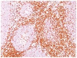
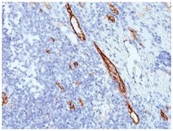
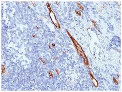
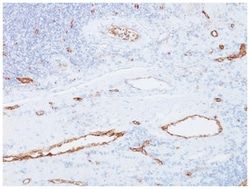
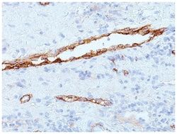
Von Willebrand Factor (vWF) is a multimeric glycoprotein that is found in endothelial cells, plasma and platelets. It acts as a carrier protein for Factor VIII and promotes platelet adhesion and aggregation. vWF undergoes a variety of posttranslational modifications that influence the affinity and availability for Factor VIII, including cleavage of the propeptide and formation of N-terminal disulfide bonds. This antibody helps to establish the endothelial nature of some lesions of disputed histogenesis, e.g. Kaposis sarcoma and cardiac myxoma. It is widely used for differentiating vascular lesions from those of other tissue differentiation within a panel of other vascular markers although not all tumors of endothelial differentiation contain this antigen.
Related Products


Description
- Von Willebrand Factor Monoclonal specifically detects Von Willebrand Factor in Human samples
- It is validated for Western Blot, Flow Cytometry, Immunohistochemistry, Immunocytochemistry/Immunofluorescence, Immunoprecipitation, Immunohistochemistry-Paraffin.
Compare Similar Items
Show Difference
Antigen: Von Willebrand Factor
Dilution: Western Blot 0.5-1.0ug/ml, Flow Cytometry 0.5-1ug/million cells, Immunocytochemistry/Immunofluorescence 0.5-1ug/ml, Immunoprecipitation 0.5-1ug/500ug protein lysate, Immunohistochemistry-Paraffin 0.5-1ug/ml, Immunohistochemistry-Frozen 0.5-1ug/ml
Classification: Monoclonal
Form: Purified
Regulatory Status: RUO
Target Species: Human
Gene Accession No.: P04275
Gene ID (Entrez): 7450
Immunogen: Recombinant human vWF fragment (3E2D10 & VWF635)
Primary or Secondary: Primary
Content And Storage: Store at 4C.
Molecular Weight of Antigen: 250 kDa
Clone: 3E2D10 + VWF635
Applications: Western Blot, Flow Cytometry, Immunocytochemistry, Immunofluorescence, Immunoprecipitation, Immunohistochemistry (Paraffin)
Conjugate: Unconjugated
Host Species: Mouse
Research Discipline: Cancer
Formulation: PBS with 0.05% BSA. with 0.05% Sodium Azide
Gene Alias: coagulation factor VIII VWF, F8, F8VWF, von Willebrand factor, VWD, vWF
Gene Symbols: VWF
Isotype: IgG
Purification Method: Protein A purified
Test Specificity: Von Willebrand Factor (vWF) is a multimeric glycoprotein that is found in endothelial cells, plasma and platelets. It acts as a carrier protein for Factor VIII and promotes platelet adhesion and aggregation. vWF undergoes a variety of posttranslational modifications that influence the affinity and availability for Factor VIII, including cleavage of the propeptide and formation of N-terminal disulfide bonds. This antibody helps to establish the endothelial nature of some lesions of disputed histogenesis, e.g. Kaposis sarcoma and cardiac myxoma. It is widely used for differentiating vascular lesions from those of other tissue differentiation within a panel of other vascular markers although not all tumors of endothelial differentiation contain this antigen.
Antigen: CD1a
Dilution: Western Blot 0.5-1ug/ml, Flow Cytometry 0.5-1ug/million cells, Immunocytochemistry/Immunofluorescence 1-2ug/ml, Immunoprecipitation 1-2ug/500ug protein, Immunohistochemistry-Paraffin 0.5-1ug/ml, Immunohistochemistry-Frozen 0.5-1ug/ml
Classification: Monoclonal
Form: Purified
Regulatory Status: RUO
Target Species: Human
Gene Accession No.: P06126
Gene ID (Entrez): 909
Immunogen: Human thymus cells
Primary or Secondary: Primary
Content And Storage: Store at 4C.
Molecular Weight of Antigen: 49 kDa
Clone: O10
Applications: Western Blot, Flow Cytometry, Immunocytochemistry, Immunofluorescence, Immunoprecipitation, Immunohistochemistry (Paraffin)
Conjugate: Unconjugated
Host Species: Mouse
Research Discipline: Dendritic Cell Markers, Immunology
Formulation: PBS with 0.05% BSA. with 0.05% Sodium Azide
Gene Alias: CD1, CD1a antigen, CD1A antigen, a polypeptide, CD1a molecule, cluster of differentiation 1 A, cortical thymocyte antigen CD1A, differentiation antigen CD1-alpha-3, epidermal dendritic cell marker CD1a, FCB6, HTA1, hTa1 thymocyte antigen, R4, T6, T-cell surface antigen T6/Leu-6, T-cell surface glycoprotein CD1a
Gene Symbols: CD1A
Isotype: IgG1 κ
Purification Method: Protein A purified
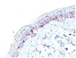
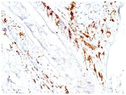
Test Specificity: At least five CD1 genes (CD1a, b, c, d, and e) are identified. CD1 proteins have been demonstrated to restrict T cell response to non-peptide lipid and glycolipid antigens and play a role in non-classical antigen presentation. CD1a is a non-polymorphic MHC Class 1 related cell surface glycoprotein, expressed in association with Beta-2 microglobulin. Anti-CD1a labels Langerhans cell histiocytosis (Histiocytosis X), extranodal histiocytic sarcoma, a subset of T-lymphoblastic lymphoma/leukemia, and interdigitating dendritic cell sarcoma of the lymph node. When combined with antibodies against TTF-1 and CD5, anti-CD1a is useful in distinguishing between pulmonary and thymic neoplasms since CD1a is consistently expressed in thymic lymphocytes in both typical and atypical thymomas, but only focally in 1/6 of thymic carcinomas and not in lymphocytes in pulmonary neoplasms. Anti-CD1a is reported to be a new marker for perivascular epithelial cell tumor (PEComa).
Antigen: CD1a
Dilution: Western Blot 0.5-1ug/ml, Flow Cytometry 0.5-1ug/million cells, Immunocytochemistry/Immunofluorescence 1-2ug/ml, Immunoprecipitation 1-2ug/500ug protein, Immunohistochemistry-Paraffin 0.5-1ug/ml, Immunohistochemistry-Frozen 0.5-1ug/ml
Classification: Monoclonal
Form: Purified
Regulatory Status: RUO
Target Species: Human
Gene Accession No.: P06126
Gene ID (Entrez): 909
Immunogen: Human thymus cells (O10); Recombinant human CD1a protein (C1A/711)
Primary or Secondary: Primary
Content And Storage: Store at 4C.
Molecular Weight of Antigen: 49 kDa
Clone: O10+C1A/711
Applications: Western Blot, Flow Cytometry, Immunocytochemistry, Immunofluorescence, Immunoprecipitation, Immunohistochemistry (Paraffin)
Conjugate: Unconjugated
Host Species: Mouse
Research Discipline: Dendritic Cell Markers, Immunology
Formulation: PBS with 0.05% BSA. with 0.05% Sodium Azide
Gene Alias: CD1, CD1a antigen, CD1A antigen, a polypeptide, CD1a molecule, cluster of differentiation 1 A, cortical thymocyte antigen CD1A, differentiation antigen CD1-alpha-3, epidermal dendritic cell marker CD1a, FCB6, HTA1, hTa1 thymocyte antigen, R4, T6, T-cell surface antigen T6/Leu-6, T-cell surface glycoprotein CD1a
Gene Symbols: CD1A
Isotype: IgG
Purification Method: Protein A purified
Test Specificity: At least five CD1 genes (CD1a, b, c, d, and e) are identified. CD1 proteins have been demonstrated to restrict T cell response to non-peptide lipid and glycolipid antigens and play a role in non-classical antigen presentation. CD1a is a non-polymorphic MHC Class 1 related cell surface glycoprotein, expressed in association with Beta-2 microglobulin. Anti-CD1a labels Langerhans cell histiocytosis (Histiocytosis X), extranodal histiocytic sarcoma, a subset of T-lymphoblastic lymphoma/leukemia, and interdigitating dendritic cell sarcoma of the lymph node. When combined with antibodies against TTF-1 and CD5, anti-CD1a is useful in distinguishing between pulmonary and thymic neoplasms since CD1a is consistently expressed in thymic lymphocytes in both typical and atypical thymomas, but only focally in 1/6 of thymic carcinomas and not in lymphocytes in pulmonary neoplasms. Anti-CD1a is reported to be a new marker for perivascular epithelial cell tumor (PEComa)