Myeloid Cell Marker Antibody (SPM298), Novus Biologicals™
Mouse Monoclonal Antibody
Manufacturer: Fischer Scientific
The price for this product is unavailable. Please request a quote
Antigen
Myeloid Cell Marker
Dilution
Flow Cytometry 0.5-1ug/million cells, Immunocytochemistry/Immunofluorescence 1-2ug/ml, Immunohistochemistry-Paraffin 0.5-1.0ug/ml, Immunohistochemistry-Frozen 0.5-1.0ug/ml
Classification
Monoclonal
Form
Purified
Regulatory Status
RUO
Formulation
PBS with 0.05% BSA. with 0.05% Sodium Azide
Immunogen
Human peripheral blood mononuclear cells
Primary or Secondary
Primary
Content And Storage
Store at 4C.
Molecular Weight of Antigen
183 kDa
Clone
SPM298
Applications
Flow Cytometry, Immunocytochemistry, Immunofluorescence, Immunohistochemistry (Paraffin), Immunohistochemistry (Frozen)
Conjugate
Unconjugated
Host Species
Mouse
Target Species
Human
Gene Accession No.
Unkonwn
Isotype
IgG1
Purification Method
Protein A purified
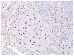
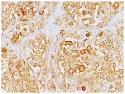
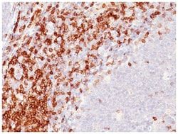
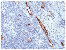
Test Specificity
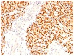
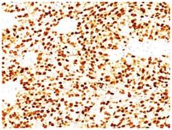
Recognizes 183kDa protein with DNA-binding characteristics, which is identified as a myeloid specific antigen. BM-1 reacts with myeloid precursor cells and granulocytes in bone marrow. Its antigen appears to be restricted to M2 and M3 acute myelogenous leukemia (AML) subtypes. Markers of myeloid cells are useful in the identification of different levels of cellular differentiation. BM-1 and BM-2 antibodies react with early precursor and mature forms of human myeloid cells. BM-1 MAb is useful in the identification of myelogenous leukemias, distinguishing granulocytic sarcomas from lymphoid malignancies and also in the study of differentiation and transformation of human myeloid cells. The biological function of this antigen is not clear, although it has been proposed that BM-1 may play a role in the differentiation of myeloid cells.
Related Products


Description
- Myeloid Cell Marker Monoclonal specifically detects Myeloid Cell Marker in Human samples
- It is validated for Flow Cytometry, Immunohistochemistry, Immunocytochemistry/Immunofluorescence, Immunohistochemistry-Paraffin.
Compare Similar Items
Show Difference
Antigen: Myeloid Cell Marker
Dilution: Flow Cytometry 0.5-1ug/million cells, Immunocytochemistry/Immunofluorescence 1-2ug/ml, Immunohistochemistry-Paraffin 0.5-1.0ug/ml, Immunohistochemistry-Frozen 0.5-1.0ug/ml
Classification: Monoclonal
Form: Purified
Regulatory Status: RUO
Formulation: PBS with 0.05% BSA. with 0.05% Sodium Azide
Immunogen: Human peripheral blood mononuclear cells
Primary or Secondary: Primary
Content And Storage: Store at 4C.
Molecular Weight of Antigen: 183 kDa
Clone: SPM298
Applications: Flow Cytometry, Immunocytochemistry, Immunofluorescence, Immunohistochemistry (Paraffin), Immunohistochemistry (Frozen)
Conjugate: Unconjugated
Host Species: Mouse
Target Species: Human
Gene Accession No.: Unkonwn
Isotype: IgG1
Purification Method: Protein A purified
Test Specificity: Recognizes 183kDa protein with DNA-binding characteristics, which is identified as a myeloid specific antigen. BM-1 reacts with myeloid precursor cells and granulocytes in bone marrow. Its antigen appears to be restricted to M2 and M3 acute myelogenous leukemia (AML) subtypes. Markers of myeloid cells are useful in the identification of different levels of cellular differentiation. BM-1 and BM-2 antibodies react with early precursor and mature forms of human myeloid cells. BM-1 MAb is useful in the identification of myelogenous leukemias, distinguishing granulocytic sarcomas from lymphoid malignancies and also in the study of differentiation and transformation of human myeloid cells. The biological function of this antigen is not clear, although it has been proposed that BM-1 may play a role in the differentiation of myeloid cells.
Antigen: MyoD
Dilution: Western Blot 0.25-0.5ug/ml, Flow Cytometry 0.5-1ug/million cells, Immunocytochemistry/Immunofluorescence 0.5-1ug/ml, Immunoprecipitation 0.5-1ug/500ug protein lysate, Immunohistochemistry-Paraffin 0.5-1.0ug/ml, Immunohistochemistry-Frozen 0.5-1.0ug/ml
Classification: Monoclonal
Form: Purified
Regulatory Status: RUO
Formulation: PBS with 0.05% BSA. with 0.05% Sodium Azide
Immunogen: Recombinant mouse MyoD1 protein
Primary or Secondary: Primary
Content And Storage: Store at 4C.
Molecular Weight of Antigen: 45 kDa
Clone: SPM427
Applications: Western Blot, Flow Cytometry, Immunocytochemistry, Immunofluorescence, Immunoprecipitation, Immunohistochemistry (Paraffin)
Conjugate: Unconjugated
Host Species: Mouse
Target Species: Human, Mouse, Rat, Chicken
Gene Accession No.: P15172
Isotype: IgG1 κ
Purification Method: Protein A purified
Test Specificity: Recognizes a phosphor-protein of 45kDa, identified as MyoD1. The epitope of this MAb maps between amino acid 180-189 in the C-terminal of mouse MyoD1 protein. It does not cross react with myogenin, Myf5, or Myf6. Antibody to MyoD1 labels the nuclei of myoblasts in developing muscle tissues. MyoD1 is not detected in normal adult tissue, but is highly expressed in the tumor cell nuclei of rhabdomyosarcomas. Occasionally nuclear expression of MyoD1 is seen in ectomesenchymoma and a subset of Wilms tumors. Weak cytoplasmic staining is observed in several non-muscle tissues, including glandular epithelium and also in rhabdomyosarcomas, neuroblastomas, Ewings sarcomas and alveolar soft part sarcomas.
Antigen: MyoD
Dilution: Flow Cytometry 0.5-1ug/million cells, Immunocytochemistry/Immunofluorescence 0.5-1ug/ml, Immunohistochemistry-Paraffin 0.5-1.0ug/ml
Classification: Monoclonal
Form: Purified
Regulatory Status: RUO
Formulation: PBS with 0.05% BSA. with 0.05% Sodium Azide
Immunogen: Recombinant human MyoD1 protein
Primary or Secondary: Primary
Content And Storage: Store at 4C.
Molecular Weight of Antigen: 45 kDa
Clone: MYD712
Applications: Flow Cytometry, Immunocytochemistry, Immunofluorescence, Immunohistochemistry (Paraffin)
Conjugate: Unconjugated
Host Species: Mouse
Target Species: Human
Gene Accession No.: P15172
Isotype: IgG1 κ
Purification Method: Protein A purified
Test Specificity: Recognizes a phosphor-protein of 45kDa, identified as MyoD1. This MAb does not cross react with myogenin, Myf5, or Myf6. Antibody to MyoD1 labels the nuclei of myoblasts in developing muscle tissues. MyoD1 is not detected in normal adult tissue, but is highly expressed in the tumor cell nuclei of rhabdomyosarcomas. Occasionally nuclear expression of MyoD1 is seen in ectomesenchymoma and a subset of Wilm s tumors. Weak cytoplasmic staining is observed in several non-muscle tissues, including glandular epithelium and also in rhabdomyosarcomas, neuroblastomas, Ewing s sarcomas and alveolar soft part sarcomas.