Bcl-xL, Clone: BX006 + 2H12, Novus Biologicals™
Mouse Monoclonal Antibody
Manufacturer: Fischer Scientific
The price for this product is unavailable. Please request a quote
Antigen
Bcl-xL
Dilution
Western Blot 0.5-1ug/ml, Flow Cytometry 0.5-1ug/million cells, Immunocytochemistry/Immunofluorescence 0.5-1ug/ml, Immunoprecipitation 0.5-1ug/500ug protein lysate, Immunohistochemistry-Paraffin 0.5-1ug/ml, Immunohistochemistry-Frozen 0.5-1ug/ml
Classification
Monoclonal
Form
Purified
Regulatory Status
RUO
Target Species
Human, Mouse, Rat, Porcine
Gene Accession No.
Q07817
Gene ID (Entrez)
598
Immunogen
A synthetic peptide, aa 3-14 (Cys-QSNRELVVDFLS) of human Bcl-X protein (BX006 & 2H12)
Primary or Secondary
Primary
Content And Storage
Store at 4C.
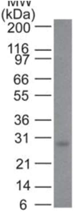
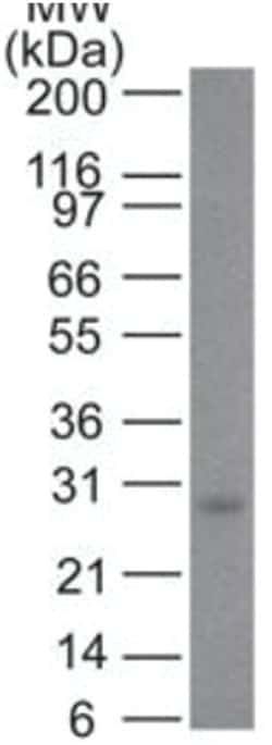
Molecular Weight of Antigen
27 kDa
Clone
BX006 + 2H12
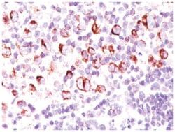
Applications
Western Blot, Flow Cytometry, Immunocytochemistry, Immunofluorescence, Immunoprecipitation, Immunohistochemistry (Paraffin)
Conjugate
Unconjugated
Host Species
Mouse
Research Discipline
Apoptosis, Cancer, Tumor Suppressors
Formulation
PBS with 0.05% BSA. with 0.05% Sodium Azide
Gene Alias
Apoptosis regulator Bcl-X, bcl2-L-1, BCL2-like 1, bcl-2-like protein 1, BCLXBCL2LBcl-X, bcl-xL, BCLXL, BCL-XL/S, bcl-xS, BCLXS, DKFZp781P2092
Gene Symbols
BCL2L1
Isotype
IgG
Purification Method
Protein A purified
Test Specificity
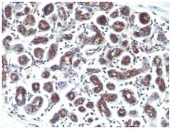
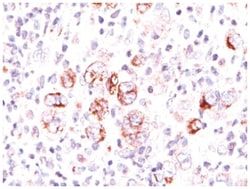
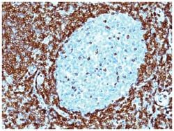
Recognizes a protein of 27kDa, identified as the Bcl-X protein. This MAb shows no cross-reaction with Bcl-2 or Bax protein. Bcl-X has two isoforms, Bcl-XL (long), a 241 amino acid protein which suppresses cell death. And Bcl-XS (short), a 178 amino acid protein lacking a 63 amino acid domain which functions as a dominant inhibitor of Bcl-2. This MAb reacts with both Bcl-XS and Bcl-XL proteins.
Related Products

Description
- Bcl-xL Monoclonal specifically detects Bcl-xL in Human, Mouse, Rat, Porcine samples
- It is validated for Western Blot, Immunohistochemistry, Immunohistochemistry-Paraffin.
Compare Similar Items
Show Difference
Antigen: Bcl-xL
Dilution: Western Blot 0.5-1ug/ml, Flow Cytometry 0.5-1ug/million cells, Immunocytochemistry/Immunofluorescence 0.5-1ug/ml, Immunoprecipitation 0.5-1ug/500ug protein lysate, Immunohistochemistry-Paraffin 0.5-1ug/ml, Immunohistochemistry-Frozen 0.5-1ug/ml
Classification: Monoclonal
Form: Purified
Regulatory Status: RUO
Target Species: Human, Mouse, Rat, Porcine
Gene Accession No.: Q07817
Gene ID (Entrez): 598
Immunogen: A synthetic peptide, aa 3-14 (Cys-QSNRELVVDFLS) of human Bcl-X protein (BX006 & 2H12)
Primary or Secondary: Primary
Content And Storage: Store at 4C.
Molecular Weight of Antigen: 27 kDa
Clone: BX006 + 2H12
Applications: Western Blot, Flow Cytometry, Immunocytochemistry, Immunofluorescence, Immunoprecipitation, Immunohistochemistry (Paraffin)
Conjugate: Unconjugated
Host Species: Mouse
Research Discipline: Apoptosis, Cancer, Tumor Suppressors
Formulation: PBS with 0.05% BSA. with 0.05% Sodium Azide
Gene Alias: Apoptosis regulator Bcl-X, bcl2-L-1, BCL2-like 1, bcl-2-like protein 1, BCLXBCL2LBcl-X, bcl-xL, BCLXL, BCL-XL/S, bcl-xS, BCLXS, DKFZp781P2092
Gene Symbols: BCL2L1
Isotype: IgG
Purification Method: Protein A purified
Test Specificity: Recognizes a protein of 27kDa, identified as the Bcl-X protein. This MAb shows no cross-reaction with Bcl-2 or Bax protein. Bcl-X has two isoforms, Bcl-XL (long), a 241 amino acid protein which suppresses cell death. And Bcl-XS (short), a 178 amino acid protein lacking a 63 amino acid domain which functions as a dominant inhibitor of Bcl-2. This MAb reacts with both Bcl-XS and Bcl-XL proteins.
Antigen: CD98
Dilution: Western Blot 0.5-1.0ug/ml, Flow Cytometry 0.5-1ug/million cells, Immunocytochemistry/Immunofluorescence 0.5-1.0ug/ml, Immunoprecipitation 1-2ug/500ug protein, Immunohistochemistry-Frozen 0.5-1ug/ml, SDS-Page
Classification: Monoclonal
Form: Purified
Regulatory Status: RUO
Target Species: Human
Gene Accession No.: P08195
Gene ID (Entrez): 6520
Immunogen: Molt 13 T cell line
Primary or Secondary: Primary
Content And Storage: Store at 4C.
Molecular Weight of Antigen: __
Clone: UM7F8
Applications: Western Blot, Flow Cytometry, Immunocytochemistry, Immunofluorescence, Immunoprecipitation, Immunohistochemistry (Frozen)
Conjugate: Unconjugated
Host Species: Mouse
Research Discipline: Cell Biology, Cellular Markers, Immunology, Lipid and Metabolism, Plasma Membrane Markers, Signal Transduction
Formulation: PBS with 0.05% BSA. with 0.05% Sodium Azide
Gene Alias: 4F2, 4F2hc, 4T2HC, antigen identified by monoclonal antibodies 4F2, TRA1.10, TROP4, and T43,4F2HC, CD98, CD98 antigen, CD98 heavy chain, CD98HC, heavy chain, Lymphocyte activation antigen 4F2 large subunit, MDU1antigen defined by monoclonal 4F2, heavy chain, monoclonal 44D7, NACAE4F2 cell-surface antigen heavy chain, solute carrier family 3 (activators of dibasic and neutral amino acidtransport), member 2,4F2 heavy chain antigen
Gene Symbols: SLC3A2
Isotype: IgG1
Purification Method: Protein G purified
Test Specificity: CD98 exits as a heterodimer containing a disulphide-linked glycosylated heavy chain and a non-glycosylated light chain. It is a member of the solute carrier family and encodes a cell surface, transmembrane protein. The protein exists as the heavy chain of a heterodimer, covalently bound through disulfide bonds to one of several possible light chains. The encoded transporter plays a role in regulation of intracellular calcium levels and transports L-type amino acids. Alternatively spliced transcript variants, encoding different isoforms, have been characterized.
Antigen: SUMO2/3
Dilution: Western Blot 0.5-1ug/ml, Flow Cytometry 0.5-1ug/million cells, Immunocytochemistry/Immunofluorescence 0.5-1ug/ml, Immunoprecipitation 0.5-1ug/500ug protein lysate, Immunohistochemistry-Paraffin 0.5-1ug/ml, Immunohistochemistry-Frozen 0.5-1ug/ml
Classification: Monoclonal
Form: Purified
Regulatory Status: RUO
Target Species: Human
Gene Accession No.: P61956
Gene ID (Entrez): 6613
Immunogen: Recombinant human SUMO2 protein
Primary or Secondary: Primary
Content And Storage: Store at 4C.
Molecular Weight of Antigen: __
Clone: SM23/496
Applications: Western Blot, Flow Cytometry, Immunocytochemistry, Immunofluorescence, Immunoprecipitation, Immunohistochemistry (Paraffin)
Conjugate: Unconjugated
Host Species: Mouse
Research Discipline: __
Formulation: PBS with 0.05% BSA. with 0.05% Sodium Azide
Gene Alias: __
Gene Symbols: SUMO2
Isotype: IgG1 κ
Purification Method: Protein A purified
Test Specificity: This MAb reacts with both SUMO-2 and SUMO-3. The small ubiquitin-related modifier (SUMO) proteins, which include SUMO-1, 2 and 3, belong to the ubiquitin-like protein family. Like ubiquitin, the SUMO proteins are synthesized as precursor proteins that undergo processing before conjugation to target proteins. Also, both utilize the E1, E2 and E3 cascade enzymes for conjugation. However, SUMO and ubiquitin differ with respect to targeting. Ubiquitination predominantly targets proteins for degradation, whereas sumoylation targets proteins to a variety of cellular processing, including nuclear transport, transcriptional regulation, apoptosis and protein stability. The unconjugated SUMO-1, 2 and 3 proteins localize to the nuclear membrane, nuclear bodies and cytoplasm, respectively. SUMO-1 utilizes Ubc9 for conjugation to several target proteins, which include MDM2, p53, PML and RanGap1. SUMO-2 and 3 contribute to a greater percentage of protein modification than does SUMO-1 and unlike SUMO