HLA DRB1 Antibody (HLA-DRB/1067) - Azide and BSA Free, Novus Biologicals™
Mouse Monoclonal Antibody
Manufacturer: Fischer Scientific
The price for this product is unavailable. Please request a quote
Antigen
HLA DRB1
Concentration
1.0 mg/mL
Applications
Western Blot, Flow Cytometry, Immunohistochemistry (Paraffin), Immunofluorescence, CyTOF
Conjugate
Unconjugated
Host Species
Mouse
Research Discipline
Asthma, Immunology
Formulation
PBS with No Preservative
Gene ID (Entrez)
3123
Isotype
IgG2b κ
Purification Method
Protein A or G purified
Test Specificity
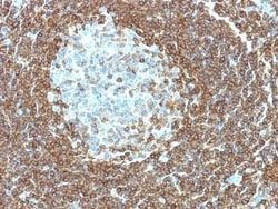
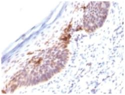
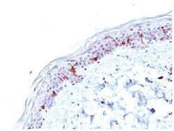
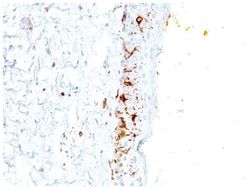
This MAb reacts with the beta-chain of HLA-DRB1 antigen, a member of MHC class II molecules. It does not cross react with HLA-DP and HLA-DQ. Its epitope is different from that of MAb L243. HLA-DR is a heterodimeric cell surface glycoprotein comprised of a 36kDa alpha (heavy) chain and a 28kDa beta (light) chain. It is expressed on B-cells, activated T-cells, monocytes/macrophages, dendritic cells and other non-professional APCs. In conjunction with the CD3/TCR complex and CD4 molecules, HLA-DR is critical for efficient peptide presentation to CD4+ T cells. It is an excellent histiocytic marker in paraffin sections producing intense cytoplasmic staining. True histiocytic neoplasms are similarly positive. HLA-DR antigens also occur on a variety of epithelial cells and their corresponding neoplastic counterparts. Loss of HLA-DR expression is related to tumor microenvironment and predicts adverse outcome in diffuse large B-cell lymphoma.
Clone
HLA-DRB/1067
Dilution
Western Blot : 0.5 - 1.0 ug/ml, Flow Cytometry : 0.5 - 1 ug/million cells in 0.1 ml, Immunohistochemistry-Paraffin : 0.25 - 0.5 ug/ml, Immunofluorescence : 0.5 - 1.0 ug/ml, CyTOF-ready
Classification
Monoclonal
Form
Purified
Regulatory Status
RUO
Target Species
Human, Monkey
Gene Alias
Clone P2-beta-3, DR1, DR-1, DR12, DR-12, DR13, DR-13, DR14, DR-14, DR16, DR-16, DR4, DR-4, DR5, DR-5, DR7, DR-7, DR8, DR-8, DR9, DR-9, DRB1, DRw10, DRw11, DRw8, DW2.2/DR2.2, FLJ75017, FLJ76359, HLA class II antigen beta chain, HLA class II histocompatibility antigen, DR-1 beta chain, HLA-DR1B, HLA-DRB, HLA-DRB1*, HLA-DRB2, HLA-DR-beta 1, human leucocyte antigen DRB1, leucocyte antigen DR beta 1 chain, leucocyte antigen DRB1, lymphocyte antigen DRB1, major histocompatibility complex, class II, DR beta 1, MHC class II antigen DRB1*1, MHC class II antigen DRB1*10, MHC class II antigen DRB1*11, MHC class II antigen DRB1*12, MHC class II antigen DRB1*13, MHC class II antigen DRB1*14, MHC class II antigen DRB1*15, MHC class II antigen DRB1*16, MHC class II antigen DRB1*3, MHC class II antigen DRB1*4, MHC class II antigen DRB1*7, MHC class II antigen DRB1*8, MHC class II antigen DRB1*9, MHC class II antigen HLA-DR13, MHC class II HLA-DR beta 1 chain, MHC class II HLA-DR-beta cell surface gly
Immunogen
Activated human peripheral blood mononuclear cells
Primary or Secondary
Primary
Content And Storage
Store at 4C short term. Aliquot and store at -20C long term. Avoid freeze-thaw cycles.
Related Products

Description
- HLA DRB1 Monoclonal specifically detects HLA DRB1 in Human, Monkey samples
- It is validated for Western Blot, Flow Cytometry, Immunohistochemistry, Immunocytochemistry/Immunofluorescence, Immunohistochemistry-Paraffin, Immunofluorescence, CyTOF-ready.
Compare Similar Items
Show Difference
Antigen: HLA DRB1
Concentration: 1.0 mg/mL
Applications: Western Blot, Flow Cytometry, Immunohistochemistry (Paraffin), Immunofluorescence, CyTOF
Conjugate: Unconjugated
Host Species: Mouse
Research Discipline: Asthma, Immunology
Formulation: PBS with No Preservative
Gene ID (Entrez): 3123
Isotype: IgG2b κ
Purification Method: Protein A or G purified
Test Specificity: This MAb reacts with the beta-chain of HLA-DRB1 antigen, a member of MHC class II molecules. It does not cross react with HLA-DP and HLA-DQ. Its epitope is different from that of MAb L243. HLA-DR is a heterodimeric cell surface glycoprotein comprised of a 36kDa alpha (heavy) chain and a 28kDa beta (light) chain. It is expressed on B-cells, activated T-cells, monocytes/macrophages, dendritic cells and other non-professional APCs. In conjunction with the CD3/TCR complex and CD4 molecules, HLA-DR is critical for efficient peptide presentation to CD4+ T cells. It is an excellent histiocytic marker in paraffin sections producing intense cytoplasmic staining. True histiocytic neoplasms are similarly positive. HLA-DR antigens also occur on a variety of epithelial cells and their corresponding neoplastic counterparts. Loss of HLA-DR expression is related to tumor microenvironment and predicts adverse outcome in diffuse large B-cell lymphoma.
Clone: HLA-DRB/1067
Dilution: Western Blot : 0.5 - 1.0 ug/ml, Flow Cytometry : 0.5 - 1 ug/million cells in 0.1 ml, Immunohistochemistry-Paraffin : 0.25 - 0.5 ug/ml, Immunofluorescence : 0.5 - 1.0 ug/ml, CyTOF-ready
Classification: Monoclonal
Form: Purified
Regulatory Status: RUO
Target Species: Human, Monkey
Gene Alias: Clone P2-beta-3, DR1, DR-1, DR12, DR-12, DR13, DR-13, DR14, DR-14, DR16, DR-16, DR4, DR-4, DR5, DR-5, DR7, DR-7, DR8, DR-8, DR9, DR-9, DRB1, DRw10, DRw11, DRw8, DW2.2/DR2.2, FLJ75017, FLJ76359, HLA class II antigen beta chain, HLA class II histocompatibility antigen, DR-1 beta chain, HLA-DR1B, HLA-DRB, HLA-DRB1*, HLA-DRB2, HLA-DR-beta 1, human leucocyte antigen DRB1, leucocyte antigen DR beta 1 chain, leucocyte antigen DRB1, lymphocyte antigen DRB1, major histocompatibility complex, class II, DR beta 1, MHC class II antigen DRB1*1, MHC class II antigen DRB1*10, MHC class II antigen DRB1*11, MHC class II antigen DRB1*12, MHC class II antigen DRB1*13, MHC class II antigen DRB1*14, MHC class II antigen DRB1*15, MHC class II antigen DRB1*16, MHC class II antigen DRB1*3, MHC class II antigen DRB1*4, MHC class II antigen DRB1*7, MHC class II antigen DRB1*8, MHC class II antigen DRB1*9, MHC class II antigen HLA-DR13, MHC class II HLA-DR beta 1 chain, MHC class II HLA-DR-beta cell surface gly
Immunogen: Activated human peripheral blood mononuclear cells
Primary or Secondary: Primary
Content And Storage: Store at 4C short term. Aliquot and store at -20C long term. Avoid freeze-thaw cycles.
Antigen: HLA DRB1
Concentration: 1.0 mg/mL
Applications: Western Blot, Flow Cytometry, Immunohistochemistry (Paraffin), Immunofluorescence, CyTOF
Conjugate: Unconjugated
Host Species: Mouse
Research Discipline: Asthma, Immunology
Formulation: PBS with No Preservative
Gene ID (Entrez): 3123
Isotype: IgG
Purification Method: Protein A or G purified
Test Specificity: This MAb reacts with the beta-chain of HLA-DRB1 antigen, a member of MHC class II molecules. It does not cross react with HLA-DP and HLA-DQ. HLA-DR is a heterodimeric cell surface glycoprotein comprised of a 36kDa alpha (heavy) chain and a 28kDa beta (light) chain. It is expressed on B-cells, activated T-cells, monocytes/macrophages, dendritic cells and other non-professional APCs. In conjunction with the CD3/TCR complex and CD4 molecules, HLA-DR is critical for efficient peptide presentation to CD4+ T cells. It is an excellent histiocytic marker in paraffin sections producing intense cytoplasmic staining. True histiocytic neoplasms are similarly positive. HLA-DR antigens also occur on a variety of epithelial cells and their corresponding neoplastic counterparts. Loss of HLA-DR expression is related to tumor microenvironment and predicts adverse outcome in diffuse large B-cell lymphoma.
Clone: LN-3 + HLA-DRB/1067
Dilution: Western Blot : 0.5 - 1.0 ug/ml, Flow Cytometry : 0.5 - 1 ug/million cells in 0.1 ml, Immunohistochemistry-Paraffin : 0.25 - 0.5 ug/ml, Immunofluorescence : 0.5 - 1.0 ug/ml, CyTOF-ready
Classification: Monoclonal
Form: Purified
Regulatory Status: RUO
Target Species: Human, Monkey
Gene Alias: Clone P2-beta-3, DR1, DR-1, DR12, DR-12, DR13, DR-13, DR14, DR-14, DR16, DR-16, DR4, DR-4, DR5, DR-5, DR7, DR-7, DR8, DR-8, DR9, DR-9, DRB1, DRw10, DRw11, DRw8, DW2.2/DR2.2, FLJ75017, FLJ76359, HLA class II antigen beta chain, HLA class II histocompatibility antigen, DR-1 beta chain, HLA-DR1B, HLA-DRB, HLA-DRB1*, HLA-DRB2, HLA-DR-beta 1, human leucocyte antigen DRB1, leucocyte antigen DR beta 1 chain, leucocyte antigen DRB1, lymphocyte antigen DRB1, major histocompatibility complex, class II, DR beta 1, MHC class II antigen DRB1*1, MHC class II antigen DRB1*10, MHC class II antigen DRB1*11, MHC class II antigen DRB1*12, MHC class II antigen DRB1*13, MHC class II antigen DRB1*14, MHC class II antigen DRB1*15, MHC class II antigen DRB1*16, MHC class II antigen DRB1*3, MHC class II antigen DRB1*4, MHC class II antigen DRB1*7, MHC class II antigen DRB1*8, MHC class II antigen DRB1*9, MHC class II antigen HLA-DR13, MHC class II HLA-DR beta 1 chain, MHC class II HLA-DR-beta cell surface gly
Immunogen: Activated human peripheral blood mononuclear cells (LN-3 and HLA-DRB/1067)
Primary or Secondary: Primary
Content And Storage: Store at 4C short term. Aliquot and store at -20C long term. Avoid freeze-thaw cycles.
Antigen: HLA A
Concentration: 1.0 mg/mL
Applications: Flow Cytometry, Immunohistochemistry (Paraffin), Immunofluorescence, CyTOF
Conjugate: Unconjugated
Host Species: Mouse
Research Discipline: Adaptive Immunity, Cell Biology, Immunology
Formulation: PBS with No Preservative
Gene ID (Entrez): 3105
Isotype: IgG2a κ
Purification Method: Protein A or G purified
Test Specificity: This MAb reacts with cells bearing HLA-A25 or HLA-Aw32 antigens. In addition, a reaction was observed with a cell of phenotype A2, Aw31; B17, Bw49. HLA-A, with HLA-B and HLA-C, belongs to major histocompatibility complex (MHC) class I antigens and expresses constitutively on all nucleated cells. HLA system comprises closely linked genes controlling highly polymorphic proteins involved in the presentation of peptides to the T-cell receptor, inhibition of NK cell cytotoxicity, and rejection of tissue allotransplantation. Specific alleles at HLA loci are associated with diseases. This MAb is specifically applicable for typing peripheral T cells for the antigens HLA-A25 and HLA-Aw32.
Clone: CATA-1
Dilution: Flow Cytometry : 0.5 - 1 ug/million cells in 0.1 ml, Immunohistochemistry-Paraffin : 0.5 - 1.0 ug/ml, Immunofluorescence : 0.5 - 1.0 ug/ml, CyTOF-ready
Classification: Monoclonal
Form: Purified
Regulatory Status: RUO
Target Species: Human
Gene Alias: A-10 alpha chain, FLJ26655, HLA class I histocompatibility antigen, A-1 alpha chain, HLA class I histocompatibility antigen, A-28 alpha chain, HLA class I histocompatibility antigen, A-9 alpha chain, HLAA, major histocompatibility complex, class I, A, MHC class I antigen A*1, MHC class I antigen A*11, MHC class I antigen A*80
Immunogen: Normal human peripheral blood lymphocytes of phenotype A1, Aw32, B7, B37, Cw-, Cw-, DR2, DRw10
Primary or Secondary: Primary
Content And Storage: Store at 4C short term. Aliquot and store at -20C long term. Avoid freeze-thaw cycles.