Cytokeratin 14 Antibody (KRT14/532) - Azide and BSA Free, Novus Biologicals™
Mouse Monoclonal Antibody
Manufacturer: Fischer Scientific
The price for this product is unavailable. Please request a quote
Antigen
Cytokeratin 14
Concentration
1.0 mg/mL
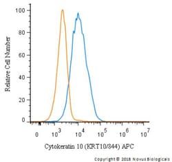
Applications
Flow Cytometry, Immunohistochemistry (Paraffin), Immunofluorescence, CyTOF
Conjugate
Unconjugated
Host Species
Mouse
Research Discipline
Cytoskeleton Markers
Formulation
PBS with No Preservative
Gene ID (Entrez)
3861
Immunogen
Recombinant full-length human KRT14 protein
Primary or Secondary
Primary
Content And Storage
Store at 4C short term. Aliquot and store at -20C long term. Avoid freeze-thaw cycles.
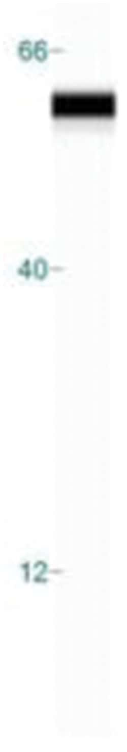
Molecular Weight of Antigen
50 kDa
Clone
KRT14/532
Dilution
Flow Cytometry : 0.5 - 1 ug/million cells in 0.1 ml, Immunohistochemistry-Paraffin : 0.5 - 1.0 ug/ml, Immunofluorescence : 0.5 - 1.0 ug/ml, CyTOF-ready
Classification
Monoclonal
Form
Purified
Regulatory Status
RUO
Target Species
Human, Mouse, Rat
Gene Alias
CK14, CK-14, cytokeratin 14, cytokeratin-14, EBS3, EBS4, K14, keratin 14, keratin 14 (epidermolysis bullosa simplex, Dowling-Meara, Koebner), keratin, type I cytoskeletal 14, Keratin-14, NFJ
Gene Symbols
KRT14
Isotype
IgG3
Purification Method
Protein A or G purified
Test Specificity
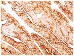
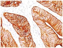
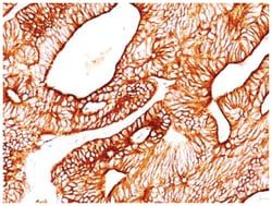
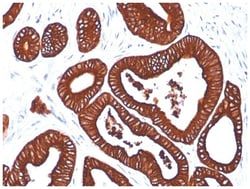
Cytokeratin 14 (CK14) belongs to the type I (or A or acidic) subfamily of low molecular weight keratins and exists in combination with keratin 5 (type II or B or basic). CK14 is found in basal cells of squamous epithelia, some glandular epithelia, myoepithelium, and mesothelial cells. Anti-CK14 is useful in differentiating squamous cell carcinomas from poorly differentiated epithelial tumors. Anti-CK14 is one of the specific basal markers for distinguishing between basal and non-basal subtypes of breast carcinomas. Anti-CK14 is also a good marker for differentiation of intraductal from invasive salivary duct carcinoma by the positive staining of basal cells surrounding the in-situ neoplasm as well as for differentiation of benign prostate from prostate carcinoma. Furthermore, this antibody has been useful in separating oncocytic tumors of the kidney from its renal mimics, and in identifying metaplastic carcinomas of the breast.
Related Products


Description
- Cytokeratin 14 Monoclonal specifically detects Cytokeratin 14 in Human, Mouse, Rat samples
- It is validated for Flow Cytometry, Immunohistochemistry, Immunocytochemistry/Immunofluorescence, Immunohistochemistry-Paraffin, Immunofluorescence, CyTOF-ready.
Compare Similar Items
Show Difference

Antigen: Cytokeratin 14
Concentration: 1.0 mg/mL
Applications: Flow Cytometry, Immunohistochemistry (Paraffin), Immunofluorescence, CyTOF
Conjugate: Unconjugated
Host Species: Mouse
Research Discipline: Cytoskeleton Markers
Formulation: PBS with No Preservative
Gene ID (Entrez): 3861
Immunogen: Recombinant full-length human KRT14 protein
Primary or Secondary: Primary
Content And Storage: Store at 4C short term. Aliquot and store at -20C long term. Avoid freeze-thaw cycles.
Molecular Weight of Antigen: 50 kDa
Clone: KRT14/532
Dilution: Flow Cytometry : 0.5 - 1 ug/million cells in 0.1 ml, Immunohistochemistry-Paraffin : 0.5 - 1.0 ug/ml, Immunofluorescence : 0.5 - 1.0 ug/ml, CyTOF-ready
Classification: Monoclonal
Form: Purified
Regulatory Status: RUO
Target Species: Human, Mouse, Rat
Gene Alias: CK14, CK-14, cytokeratin 14, cytokeratin-14, EBS3, EBS4, K14, keratin 14, keratin 14 (epidermolysis bullosa simplex, Dowling-Meara, Koebner), keratin, type I cytoskeletal 14, Keratin-14, NFJ
Gene Symbols: KRT14
Isotype: IgG3
Purification Method: Protein A or G purified
Test Specificity: Cytokeratin 14 (CK14) belongs to the type I (or A or acidic) subfamily of low molecular weight keratins and exists in combination with keratin 5 (type II or B or basic). CK14 is found in basal cells of squamous epithelia, some glandular epithelia, myoepithelium, and mesothelial cells. Anti-CK14 is useful in differentiating squamous cell carcinomas from poorly differentiated epithelial tumors. Anti-CK14 is one of the specific basal markers for distinguishing between basal and non-basal subtypes of breast carcinomas. Anti-CK14 is also a good marker for differentiation of intraductal from invasive salivary duct carcinoma by the positive staining of basal cells surrounding the in-situ neoplasm as well as for differentiation of benign prostate from prostate carcinoma. Furthermore, this antibody has been useful in separating oncocytic tumors of the kidney from its renal mimics, and in identifying metaplastic carcinomas of the breast.
Antigen: IgM
Concentration: 1.0 mg/mL
Applications: Flow Cytometry, Immunohistochemistry (Paraffin), Immunofluorescence, CyTOF
Conjugate: Unconjugated
Host Species: Mouse
Research Discipline: B Cell Development and Differentiation Markers, Immunology
Formulation: PBS with No Preservative
Gene ID (Entrez): 3507
Immunogen: Heavy chain of human IgM
Primary or Secondary: Primary
Content And Storage: Store at 4C short term. Aliquot and store at -20C long term. Avoid freeze-thaw cycles.
Molecular Weight of Antigen: __
Clone: ICO-30
Dilution: Flow Cytometry : 0.5 - 1 ug/million cells in 0.1 ml, Immunohistochemistry-Paraffin : 0.5 - 1.0 ug/ml, Immunofluorescence : 0.5 - 1.0 ug/ml, CyTOF-ready
Classification: Monoclonal
Form: Purified
Regulatory Status: RUO
Target Species: Human
Gene Alias: AGM1, DKFZp686I15196, DKFZp686I15212, FLJ00385, immunoglobulin heavy constant mu, MGC104996, MGC52291, MU, VH
Gene Symbols: IGHM
Isotype: IgG1 κ
Purification Method: Protein A or G purified
Test Specificity: Recognizes a protein of 75kDa, identified as mu heavy chain of human immunoglobulins. It does not cross-react with alpha (IgA), gamma (IgG), epsilon (IgE), or delta (IgD), heavy chains, T-cells, monocytes, granulocytes, or erythrocytes. Monomeric IgM is expressed as a membrane bound antibody on the surface of B cells and as a pentamer when secreted by plasma cells. IgM antibody is prominent in early immune responses to most antigens. Aberrant levels are associated with immune deficiency states, hereditary deficiencies, myeloma, Waldenstrom's macroglobulinemia, chronic infection and hepatocellular disease. This MAb is useful in the identification of leukemias, plasmacytomas, and certain non-Hodgkin's lymphomas. The most common feature of these malignancies is the restricted expression of a single heavy chain class. Demonstration of clonality in lymphoid infiltrates indicates that the infiltrate is clonal and therefore malignant.
Antigen: IgG
Concentration: 1.0 mg/mL
Applications: Flow Cytometry, Immunohistochemistry (Paraffin), Immunofluorescence, CyTOF
Conjugate: Unconjugated
Host Species: Mouse
Research Discipline: Immunology
Formulation: PBS with No Preservative
Gene ID (Entrez): 3500
Immunogen: Purified polyclonal human Ig Gamma Chain
Primary or Secondary: Primary
Content And Storage: Store at 4C short term. Aliquot and store at -20C long term. Avoid freeze-thaw cycles.
Molecular Weight of Antigen: 75 kDa
Clone: B33/20
Dilution: Flow Cytometry : 0.5 - 1 ug/million cells in 0.1 ml, Immunohistochemistry-Paraffin : 0.5 - 1.0 ug/ml, Immunofluorescence : 0.5 - 1.0 ug/ml, CyTOF-ready
Classification: Monoclonal
Form: Purified
Regulatory Status: RUO
Target Species: Human
Gene Alias: immunoglobulin heavy constant gamma 1 (G1m marker)
Gene Symbols: IGHG
Isotype: IgG1 κ
Purification Method: Protein A or G purified
Test Specificity: Recognizes a protein of 75kDa, identified as gamma heavy chain of human immunoglobulins. Its epitope maps in CH2 domain of Fc region of IgG. It reacts with all sub-classes of gamma chain of human immunoglobulins. It does not cross-react with alpha (IgA), u (IgM), epsilon (IgE), or delta (IgD), heavy chains, T-cells, monocytes, granulocytes, or erythrocytes. This MAb is useful in the identification of leukemias, plasmacytomas, and certain non-Hodgkin's lymphomas. The most common feature of these malignancies is the restricted expression of a single heavy chain class. Demonstration of clonality in lymphoid infiltrates indicates that the infiltrate is clonal and therefore malignant.