c-Myc Antibody (9E10.3) - Azide and BSA Free, Novus Biologicals™
Mouse Monoclonal Antibody
Manufacturer: Fischer Scientific
The price for this product is unavailable. Please request a quote
Antigen
c-Myc
Concentration
1.0 mg/mL
Applications
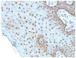
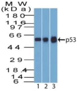
Western Blot, Flow Cytometry, Immunohistochemistry (Paraffin), Immunofluorescence, CyTOF
Conjugate
Unconjugated
Host Species
Mouse
Research Discipline
Autophagy, Cancer, Cancer Stem Cells, Cell Cycle and Replication, Chromatin Research, Core ESC Like Genes, Epitope Tags, Myc Epitope Tags, Stem Cell Markers, Transcription Factors and Regulators, Tumor Suppressors
Formulation
PBS with No Preservative
Gene ID (Entrez)
4609
Immunogen
A synthetic peptide, corresponding to aa 408-439 (AEEQKLISEEDLLRKRREQLKHKLEQLRNSCA) from C-terminus of human c-myc, coupled to KLH.
Primary or Secondary
Primary
Content And Storage
Store at 4C short term. Aliquot and store at -20C long term. Avoid freeze-thaw cycles.
Clone
9E10.3
Dilution
Simple Western : 50 ug/ml, Flow Cytometry : 0.5 - 1 ug/million cells in 0.1 ml, Immunohistochemistry-Paraffin : 1 - 2 ug/ml, Immunofluorescence : 1 - 2 ug/ml, CyTOF-ready
Classification
Monoclonal
Form
Purified
Regulatory Status
RUO
Target Species
Human, Chicken (Negative), Mouse (Negative), Rat (Negative)
Gene Alias
avian myelocytomatosis viral oncogene homolog, BHLHE39, bHLHe39MRTL, Class E basic helix-loop-helix protein 39, c-Myc, MYC, myc proto-oncogene protein, MYCC, myc-related translation/localization regulatory factor, Proto-oncogene c-Myc, Transcription factor p64, v-myc avian myelocytomatosis viral oncogene homolog, v-myc myelocytomatosis viral oncogene homolog (avian)
Gene Symbols
MYC
Isotype
IgG1 κ
Purification Method
Protein A or G purified
Test Specificity
It recognizes a transcription factor of 64-67kDa, identified as c-myc. Its epitope spans between aa 410-419 (EQKLISEEDL) which is a specific portion of an alpha helical region of human c-myc protein. This MAb shows no cross-reaction with v-myc. c-myc is involved in the control of cell proliferation and differentiation and is amplified and/or overexpressed in a variety of tumors. Over-expression of c-myc protein occurs frequently in luminal cells of prostate intraepithelial neoplasia as well as in most primary carcinomas and metastatic disease
Related Products

Description
- c-Myc Monoclonal specifically detects c-Myc in Human, Chicken (Negative), Mouse (Negative), Rat (Negative) samples
- It is validated for Simple Western, Flow Cytometry, Immunohistochemistry, Immunocytochemistry/Immunofluorescence, Immunohistochemistry-Paraffin, Immunofluorescence, CyTOF-ready.
Compare Similar Items
Show Difference
Antigen: c-Myc
Concentration: 1.0 mg/mL
Applications: Western Blot, Flow Cytometry, Immunohistochemistry (Paraffin), Immunofluorescence, CyTOF
Conjugate: Unconjugated
Host Species: Mouse
Research Discipline: Autophagy, Cancer, Cancer Stem Cells, Cell Cycle and Replication, Chromatin Research, Core ESC Like Genes, Epitope Tags, Myc Epitope Tags, Stem Cell Markers, Transcription Factors and Regulators, Tumor Suppressors
Formulation: PBS with No Preservative
Gene ID (Entrez): 4609
Immunogen: A synthetic peptide, corresponding to aa 408-439 (AEEQKLISEEDLLRKRREQLKHKLEQLRNSCA) from C-terminus of human c-myc, coupled to KLH.
Primary or Secondary: Primary
Content And Storage: Store at 4C short term. Aliquot and store at -20C long term. Avoid freeze-thaw cycles.
Clone: 9E10.3
Dilution: Simple Western : 50 ug/ml, Flow Cytometry : 0.5 - 1 ug/million cells in 0.1 ml, Immunohistochemistry-Paraffin : 1 - 2 ug/ml, Immunofluorescence : 1 - 2 ug/ml, CyTOF-ready
Classification: Monoclonal
Form: Purified
Regulatory Status: RUO
Target Species: Human, Chicken (Negative), Mouse (Negative), Rat (Negative)
Gene Alias: avian myelocytomatosis viral oncogene homolog, BHLHE39, bHLHe39MRTL, Class E basic helix-loop-helix protein 39, c-Myc, MYC, myc proto-oncogene protein, MYCC, myc-related translation/localization regulatory factor, Proto-oncogene c-Myc, Transcription factor p64, v-myc avian myelocytomatosis viral oncogene homolog, v-myc myelocytomatosis viral oncogene homolog (avian)
Gene Symbols: MYC
Isotype: IgG1 κ
Purification Method: Protein A or G purified
Test Specificity: It recognizes a transcription factor of 64-67kDa, identified as c-myc. Its epitope spans between aa 410-419 (EQKLISEEDL) which is a specific portion of an alpha helical region of human c-myc protein. This MAb shows no cross-reaction with v-myc. c-myc is involved in the control of cell proliferation and differentiation and is amplified and/or overexpressed in a variety of tumors. Over-expression of c-myc protein occurs frequently in luminal cells of prostate intraepithelial neoplasia as well as in most primary carcinomas and metastatic disease
Antigen: EGFR
Concentration: 1.0 mg/mL
Applications: Flow Cytometry, Immunohistochemistry (Paraffin), Immunofluorescence, CyTOF
Conjugate: Unconjugated
Host Species: Mouse
Research Discipline: Cancer, Cell Biology, Cell Cycle and Replication, Growth and Development, Hypoxia, Signal Transduction, Tumor Suppressors, Tyrosine Kinases
Formulation: PBS with No Preservative
Gene ID (Entrez): 1956
Immunogen: Human EGFR purified from A431 cells
Primary or Secondary: Primary
Content And Storage: Store at 4C short term. Aliquot and store at -20C long term. Avoid freeze-thaw cycles.
Clone: SPM622
Dilution: Flow Cytometry : 0.5 - 1 ug/million cells in 0.1 ml, Immunohistochemistry-Paraffin : 2 - 4 ug/ml, Immunofluorescence : 1 - 2 ug/ml, CyTOF-ready
Classification: Monoclonal
Form: Purified
Regulatory Status: RUO
Target Species: Human
Gene Alias: avian erythroblastic leukemia viral (v-erb-b) oncogene homolog, cell growth inhibiting protein 40, cell proliferation-inducing protein 61, EC 2.7.10, EC 2.7.10.1, epidermal growth factor receptor, epidermal growth factor receptor (avian erythroblastic leukemia viral (v-erb-b)oncogene homolog), ERBB, ErbB1, ERBB1PIG61, HER1, mENA, Proto-oncogene c-ErbB-1, Receptor tyrosine-protein kinase erbB-1
Gene Symbols: EGFR
Isotype: IgG1 κ
Purification Method: Protein A or G purified
Test Specificity: This MAb recognizes a protein of 170kDa, identified as EGFR. EGFR is type I receptor tyrosine kinase with sequence homology to erbB-1, -2, -3 -4 or HER-1, -2, -3 -4. It binds to Epidermal Growth Factor (EGF), Transforming Growth Factor-a (TGF-a), Heparin-binding EGF (HB-EGF), amphiregulin, Beta cellulin and epiregulin. EGFR is overexpressed in tumors of breast, brain, bladder, lung, gastric, head & neck, esophagus, cervix, vulva, ovary, and endometrium. It is predominantly present in squamous cell carcinomas.