c-Myc Antibody (MYC275), Novus Biologicals™
Mouse Monoclonal Antibody
Manufacturer: Fischer Scientific
The price for this product is unavailable. Please request a quote
Antigen
c-Myc
Dilution
Flow Cytometry 0.5-1ug/million cells, Immunohistochemistry, Immunocytochemistry/Immunofluorescence 1-2 ug/ml, Immunohistochemistry-Paraffin 1-2 ug/ml
Classification
Monoclonal
Form
Purified
Regulatory Status
RUO
Target Species
Human
Gene Accession No.
P01106
Gene ID (Entrez)
4609
Immunogen
Recombinant human c-myc protein
Primary or Secondary
Primary
Content And Storage
Store at 4C.
Clone
MYC275
Applications
Flow Cytometry, Immunohistochemistry, Immunocytochemistry, Immunofluorescence, Immunohistochemistry (Paraffin)
Conjugate
Unconjugated
Host Species
Mouse
Research Discipline
Autophagy, Cancer, Cell Cycle and Replication, Chromatin Research, Core ESC Like Genes, Epitope Tags, Myc Epitope Tags, Phospho Specific, Stem Cell Markers, Transcription Factors and Regulators, Tumor Suppressors
Formulation
PBS with 0.05% BSA. with 0.05% Sodium Azide
Gene Alias
avian myelocytomatosis viral oncogene homolog, BHLHE39, bHLHe39MRTL, Class E basic helix-loop-helix protein 39, c-Myc, MYC, myc proto-oncogene protein, MYCC, myc-related translation/localization regulatory factor, Proto-oncogene c-Myc, Transcription factor p64, v-myc avian myelocytomatosis viral oncogene homolog, v-myc myelocytomatosis viral oncogene homolog (avian)
Gene Symbols
MYC
Isotype
IgG1 κ
Purification Method
Protein A or G purified
Test Specificity
The c-Myc protein is a transcription factor, which is encoded by the c-Myc gene on human chromosome 8q24. c-Myc is commonly activated in a variety of tumor cells and plays an important role in cellular proliferation, differentiation, apoptosis and cell cycle progression. The phosphorylation of c-Myc has been investigated and previous studies have suggested a functional association between phosphorylation at Thr58/Ser62 by glycogen synthase kinase 3, cyclin dependent kinase, ERK2 and C-Jun N terminal Kinase (JNK) in cell proliferation and cell cycle regulation. Studies also have shown that c-Myc is essential for tumor cell development in vasculogenesis and angiogenesis that distribute blood throughout the cells, and which brought extensive attention in the development of new therapeutic approach for cancer treatment.
Related Products

Description
- c-Myc Monoclonal specifically detects c-Myc in Human samples
- It is validated for Flow Cytometry, Immunohistochemistry, Immunocytochemistry/Immunofluorescence, Immunohistochemistry-Paraffin.
Compare Similar Items
Show Difference
Antigen: c-Myc
Dilution: Flow Cytometry 0.5-1ug/million cells, Immunohistochemistry, Immunocytochemistry/Immunofluorescence 1-2 ug/ml, Immunohistochemistry-Paraffin 1-2 ug/ml
Classification: Monoclonal
Form: Purified
Regulatory Status: RUO
Target Species: Human
Gene Accession No.: P01106
Gene ID (Entrez): 4609
Immunogen: Recombinant human c-myc protein
Primary or Secondary: Primary
Content And Storage: Store at 4C.
Clone: MYC275
Applications: Flow Cytometry, Immunohistochemistry, Immunocytochemistry, Immunofluorescence, Immunohistochemistry (Paraffin)
Conjugate: Unconjugated
Host Species: Mouse
Research Discipline: Autophagy, Cancer, Cell Cycle and Replication, Chromatin Research, Core ESC Like Genes, Epitope Tags, Myc Epitope Tags, Phospho Specific, Stem Cell Markers, Transcription Factors and Regulators, Tumor Suppressors
Formulation: PBS with 0.05% BSA. with 0.05% Sodium Azide
Gene Alias: avian myelocytomatosis viral oncogene homolog, BHLHE39, bHLHe39MRTL, Class E basic helix-loop-helix protein 39, c-Myc, MYC, myc proto-oncogene protein, MYCC, myc-related translation/localization regulatory factor, Proto-oncogene c-Myc, Transcription factor p64, v-myc avian myelocytomatosis viral oncogene homolog, v-myc myelocytomatosis viral oncogene homolog (avian)
Gene Symbols: MYC
Isotype: IgG1 κ
Purification Method: Protein A or G purified
Test Specificity: The c-Myc protein is a transcription factor, which is encoded by the c-Myc gene on human chromosome 8q24. c-Myc is commonly activated in a variety of tumor cells and plays an important role in cellular proliferation, differentiation, apoptosis and cell cycle progression. The phosphorylation of c-Myc has been investigated and previous studies have suggested a functional association between phosphorylation at Thr58/Ser62 by glycogen synthase kinase 3, cyclin dependent kinase, ERK2 and C-Jun N terminal Kinase (JNK) in cell proliferation and cell cycle regulation. Studies also have shown that c-Myc is essential for tumor cell development in vasculogenesis and angiogenesis that distribute blood throughout the cells, and which brought extensive attention in the development of new therapeutic approach for cancer treatment.
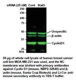
Antigen: Cyclin D1
Dilution: Western Blot 0.5-1ug/ml, Flow Cytometry 0.5-1ug/million cells, Immunocytochemistry/Immunofluorescence 1-2ug/ml, Immunoprecipitation 1 - 2ug/500ug protein lysate, Immunohistochemistry-Paraffin 0.5-1.0ug/ml
Classification: Monoclonal
Form: Purified
Regulatory Status: RUO
Target Species: Human, Mouse, Rat, Primate
Gene Accession No.: P24385, P24385, P25322, P39948
Gene ID (Entrez): 595
Immunogen: Human recombinant full length cyclin D1 protein
Primary or Secondary: Primary
Content And Storage: Store at 4C.
Clone: SPM587
Applications: Western Blot, Flow Cytometry, Immunocytochemistry, Immunofluorescence, Immunoprecipitation, Immunohistochemistry (Paraffin)
Conjugate: Unconjugated
Host Species: Mouse
Research Discipline: Cancer, Cell Cycle and Replication, Core ESC Like Genes, mTOR Pathway, Stem Cell Markers, Wnt Signaling Pathway
Formulation: PBS with 0.05% BSA. with 0.05% Sodium Azide
Gene Alias: B-cell lymphoma 1 protein, BCL-1, BCL-1 oncogene, BCL1D11S287E, cyclin D1, cyclin D1 (PRAD1: parathyroid adenomatosis 1), G1/S-specific cyclin D1, G1/S-specific cyclin-D1, PRAD1 oncogene, PRAD1B-cell CLL/lymphoma 1, U21B31
Gene Symbols: CCND1
Isotype: IgG2a κ
Purification Method: Protein A purified
Test Specificity: Recognizes a protein of 36kDa, identified as cyclin D1. Cyclin D1, one of the key cell cycle regulators, is a putative proto-oncogene overexpressed in a wide variety of human neoplasms. This antibody neutralizes the activity of cyclin D1 in vivo. About 60% of mantle cell lymphomas (MCL) contain a t(11; 14)(q13; q32) translocation resulting in over-expression of cyclin D1. This antibody is useful in identifying mantle cell lymphomas (cyclin D1 positive) from CLL/SLL and follicular lymphomas (cyclin D1 negative). Occasionally, hairy cell leukemia and plasma cell myeloma weakly express Cyclin D1.
Antigen: EGFR
Dilution: Flow Cytometry 0.5-1ug/million cells, Immunoprecipitation 0.5-1ug/500ug protein lysate
Classification: Monoclonal
Form: Purified
Regulatory Status: RUO
Target Species: Human, Mouse (Negative), Rat (Negative)
Gene Accession No.: P00533
Gene ID (Entrez): 1956
Immunogen: Microsomes from A431 cells
Primary or Secondary: Primary
Content And Storage: Store at 4C.
Clone: B1D8
Applications: Flow Cytometry, Immunoprecipitation
Conjugate: Unconjugated
Host Species: Mouse
Research Discipline: Cancer, Cell Biology, Cell Cycle and Replication, Growth and Development, Hypoxia, Phospho Specific, Signal Transduction, Tumor Suppressors, Tyrosine Kinases
Formulation: PBS with 0.05% BSA. with 0.05% Sodium Azide
Gene Alias: avian erythroblastic leukemia viral (v-erb-b) oncogene homolog, cell growth inhibiting protein 40, cell proliferation-inducing protein 61, EC 2.7.10, EC 2.7.10.1, epidermal growth factor receptor, epidermal growth factor receptor (avian erythroblastic leukemia viral (v-erb-b)oncogene homolog), ERBB, ErbB1, ERBB1PIG61, HER1, mENA, Proto-oncogene c-ErbB-1, Receptor tyrosine-protein kinase erbB-1
Gene Symbols: EGFR
Isotype: IgG2a κ
Purification Method: Protein A purified
Test Specificity: This antibody reacts with the extracellular domain of EGFR and blocks the EGF/TGF??induced activation. It also blocks tumor growth in vivo. It is excellent for purification of EGFR. EGFR is type I receptor tyrosine kinase with sequence homology to erbB-1, -2, -3 -4 or HER-1, -2, -3 -4. It binds to Epidermal Growth Factor (EGF), Transforming Growth Factor-a (TGF-a), Heparin-binding EGF (HB-EGF), amphiregulin, Beta cellulin and epiregulin.