Cadherin-16 Antibody (SPM594) - Azide and BSA Free, Novus Biologicals™
Mouse Monoclonal Antibody
Manufacturer: Fischer Scientific
The price for this product is unavailable. Please request a quote
Antigen
Cadherin-16
Concentration
1.0 mg/mL
Applications
Flow Cytometry, Immunohistochemistry (Paraffin), CyTOF
Conjugate
Unconjugated
Host Species
Mouse
Target Species
Human, Mouse, Rat, Canine, Rabbit
Gene Alias
cadherin 16, KSP-cadherin, cadherin-16, Kidney-specific cadherin, KSP-cadherin
Gene Symbols
CDH16
Isotype
IgG1 κ
Purification Method
Protein A or G purified
Test Specificity
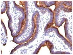
This MAb recognizes a protein of 130kDa, identified as Ksp-cadherin. Cadherins form a superfamily of related glycoproteins that mediate calcium-dependent cell adhesion and transmit signals from the extracellular matrix to the cytoplasm. Cadherins have been implicated in embryogenesis, tissue morphogenesis, tissue structure maintenance, cell polarization, neoplastic invasiveness and metastasis, and membrane transport. It is suggested that Ksp-cadherin is a marker for terminal differentiation of the basolateral membranes of renal tubular epithelial cells. Within the kidney, Ksp-Cadherin is found exclusively in the basolateral membrane of renal tubular epithelial cells and collecting duct cells, and not in glomeruli, renal interstitial cells, or blood vessels.Ksp-Cadherin has been suggested to distinguish Chromophobe Renal-Cell Carcinoma from Oncocytoma.
Clone
SPM594
Dilution
Flow Cytometry : 0.5 - 1 ug/million cells in 0.1 ml, Immunohistochemistry-Paraffin : 0.5 - 1.0 ug/ml, CyTOF-ready
Classification
Monoclonal
Form
Purified
Regulatory Status
RUO
Formulation
PBS with No Preservative
Gene ID (Entrez)
1014
Immunogen
Recombinant human CDH16 protein
Primary or Secondary
Primary
Content And Storage
Store at 4C short term. Aliquot and store at -20C long term. Avoid freeze-thaw cycles.
Molecular Weight of Antigen
130 kDa
Related Products

Description
- Cadherin-16 Monoclonal specifically detects Cadherin-16 in Human, Mouse, Rat, Canine, Rabbit samples
- It is validated for Western Blot, Immunohistochemistry, Immunohistochemistry-Paraffin, CyTOF-ready.
Compare Similar Items
Show Difference
Antigen: Cadherin-16
Concentration: 1.0 mg/mL
Applications: Flow Cytometry, Immunohistochemistry (Paraffin), CyTOF
Conjugate: Unconjugated
Host Species: Mouse
Target Species: Human, Mouse, Rat, Canine, Rabbit
Gene Alias: cadherin 16, KSP-cadherin, cadherin-16, Kidney-specific cadherin, KSP-cadherin
Gene Symbols: CDH16
Isotype: IgG1 κ
Purification Method: Protein A or G purified
Test Specificity: This MAb recognizes a protein of 130kDa, identified as Ksp-cadherin. Cadherins form a superfamily of related glycoproteins that mediate calcium-dependent cell adhesion and transmit signals from the extracellular matrix to the cytoplasm. Cadherins have been implicated in embryogenesis, tissue morphogenesis, tissue structure maintenance, cell polarization, neoplastic invasiveness and metastasis, and membrane transport. It is suggested that Ksp-cadherin is a marker for terminal differentiation of the basolateral membranes of renal tubular epithelial cells. Within the kidney, Ksp-Cadherin is found exclusively in the basolateral membrane of renal tubular epithelial cells and collecting duct cells, and not in glomeruli, renal interstitial cells, or blood vessels.Ksp-Cadherin has been suggested to distinguish Chromophobe Renal-Cell Carcinoma from Oncocytoma.
Clone: SPM594
Dilution: Flow Cytometry : 0.5 - 1 ug/million cells in 0.1 ml, Immunohistochemistry-Paraffin : 0.5 - 1.0 ug/ml, CyTOF-ready
Classification: Monoclonal
Form: Purified
Regulatory Status: RUO
Formulation: PBS with No Preservative
Gene ID (Entrez): 1014
Immunogen: Recombinant human CDH16 protein
Primary or Secondary: Primary
Content And Storage: Store at 4C short term. Aliquot and store at -20C long term. Avoid freeze-thaw cycles.
Molecular Weight of Antigen: 130 kDa
Antigen: TOX3
Concentration: 1.0 mg/mL
Applications: Flow Cytometry, Immunohistochemistry (Paraffin), Immunofluorescence, CyTOF
Conjugate: Unconjugated
Host Species: Mouse
Target Species: Human
Gene Alias: CAG trinucleotide repeat-containing gene F9 protein, CAGF9trinucleotide repeat containing 9, TNRC9, TOX high mobility group box family member 3, Trinucleotide repeat-containing gene 9 protein
Gene Symbols: TOX3
Isotype: IgG2b κ
Purification Method: Protein A or G purified
Test Specificity: It recognizes a 63kDa protein, which is identified as TOX3. It contains a high mobility group (HMG)-box, which regulates Ca2+-dependent transcription in neurons through interaction with the cAMP-response-element-binding protein (CREB). TOX3 appears to be associated with breast cancer susceptibility and is expressed downstream of a cytoprotective cascade together with CITED1, a transcriptional regulator that does not bind directly to DNA. TOX3 is predominantly expressed in the brain and forms homodimers. TOX3 overexpression protects neuronal cells from cell death caused by endoplasmic reticulum stress or BAX overexpression through the induction of anti-apoptotic transcripts and repression of pro-apoptotic transcripts.
Clone: TOX3/1123
Dilution: Flow Cytometry : 0.5 - 1 ug/million cells in 0.1 ml, Immunohistochemistry-Paraffin : 0.5 - 1.0 ug/ml, Immunofluorescence : 0.5 - 1.0 ug/ml, CyTOF-ready
Classification: Monoclonal
Form: Purified
Regulatory Status: RUO
Formulation: PBS with No Preservative
Gene ID (Entrez): 27324
Immunogen: Recombinant fragment (139 Amino acid residues around aa 200-400) of human TOX3 protein
Primary or Secondary: Primary
Content And Storage: Store at 4C short term. Aliquot and store at -20C long term. Avoid freeze-thaw cycles.
Molecular Weight of Antigen: 63 kDa
Antigen: TOX3
Concentration: 1.0 mg/mL
Applications: Western Blot, Flow Cytometry, Immunofluorescence, CyTOF
Conjugate: Unconjugated
Host Species: Mouse
Target Species: Human
Gene Alias: CAG trinucleotide repeat-containing gene F9 protein, CAGF9trinucleotide repeat containing 9, TNRC9, TOX high mobility group box family member 3, Trinucleotide repeat-containing gene 9 protein
Gene Symbols: TOX3
Isotype: IgG1 κ
Purification Method: Protein A or G purified
Test Specificity: It recognizes a 63kDa protein, which is identified as TOX3. It contains a high mobility group (HMG)-box, which regulates Ca2+-dependent transcription in neurons through interaction with the cAMP-response-element-binding protein (CREB). TOX3 appears to be associated with breast cancer susceptibility and is expressed downstream of a cytoprotective cascade together with CITED1, a transcriptional regulator that does not bind directly to DNA. TOX3 is predominantly expressed in the brain and forms homodimers. TOX3 overexpression protects neuronal cells from cell death caused by endoplasmic reticulum stress or BAX overexpression through the induction of anti-apoptotic transcripts and repression of pro-apoptotic transcripts.
Clone: TOX3/1124
Dilution: Western Blot : 0.5 - 1.0 ug/ml, Flow Cytometry : 0.5 - 1 ug/million cells in 0.1 ml, Immunofluorescence : 0.5 - 1.0 ug/ml, CyTOF-ready
Classification: Monoclonal
Form: Purified
Regulatory Status: RUO
Formulation: PBS with No Preservative
Gene ID (Entrez): 27324
Immunogen: Recombinant fragment (139 Amino acid residues around aa 200-400) of human TOX3 protein
Primary or Secondary: Primary
Content And Storage: Store at 4C short term. Aliquot and store at -20C long term. Avoid freeze-thaw cycles.
Molecular Weight of Antigen: 63 kDa