CD59 Antibody (MACIF/1193) - Azide and BSA Free, Novus Biologicals™
Mouse Monoclonal Antibody
Manufacturer: Fischer Scientific
The price for this product is unavailable. Please request a quote
Antigen
CD59
Concentration
1.0 mg/mL
Applications
Flow Cytometry, Immunohistochemistry (Paraffin), Immunofluorescence, CyTOF
Conjugate
Unconjugated
Host Species
Mouse
Research Discipline
Cell Biology, Cellular Markers, Immunology, Signal Transduction, Stem Cell Markers
Formulation
PBS with No Preservative
Gene ID (Entrez)
966
Immunogen
Recombinant full-length human CD59 protein
Primary or Secondary
Primary
Content And Storage
Store at 4C short term. Aliquot and store at -20C long term. Avoid freeze-thaw cycles.
Molecular Weight of Antigen
20 kDa
Clone
MACIF/1193
Dilution
Flow Cytometry : 0.5 - 1 ug/million cells in 0.1 ml, Immunohistochemistry-Paraffin : 1 - 2 ug/ml, Immunofluorescence : 0.5 - 1.0 ug/ml, CyTOF-ready
Classification
Monoclonal
Form
Purified
Regulatory Status
RUO
Target Species
Human, Baboon (Negative), Equine (Negative)
Gene Alias
16.3A5, 1F5, 1F5 antigen, 20 kDa homologous restriction factor, CD59 antigen, CD59 antigen p18-20 (antigen identified by monoclonal antibodies 16.3A5, EJ16, CD59 antigen, complement regulatory protein, CD59 glycoprotein, CD59 molecule, complement regulatory protein, EJ16, EJ30, EJ30, EL32 and G344), EL32, FLJ38134, FLJ92039, G344, HRF20, HRF-20, human leukocyte antigen MIC11, Ly-6-like protein, lymphocytic antigen CD59/MEM43, MACIF, MAC-inhibitory protein, MAC-IP, MEM43, MEM43 antigen, membrane attack complex (MAC) inhibition factor, Membrane attack complex inhibition factor, Membrane inhibitor of reactive lysis, MGC2354, MIC11MSK21, MIN1, MIN2, MIN3, MIRL, p18-20, protectin, surface anitgen recognized by monoclonal 16.3A5, T cell-activating protein
Gene Symbols
CD59
Isotype
IgM κ
Purification Method
Protein A or G purified
Test Specificity
Reacts with human CD59, a 20kDa glycosyl phosphatidyl-inositol (GPI)-anchored cell surface protein. CD59 regulates complement-mediated cell lysis, and it is involved in lymphocyte signal transduction. This protein is a potent inhibitor of the complement membrane attack complex, whereby it binds complement C8 and/or C9 during the assembly of this complex, thereby inhibiting the incorporation of multiple copies of C9 into the complex, which is necessary for osmolytic pore formation. CD59 is widely distributed on cells in all tissues. It inhibits formation of MAC, thus protecting cells from complement-mediated lysis. The expression of CD59 on erythrocytes is important for their survival. Genetic defects in GPI-anchor attachment, that cause a reduction or loss of CD59 and CD55 on erythrocytes produce the symptoms of the disease paroxysmal hemoglobinuria (PNH). It is useful for study on GPI-anchored proteins, PNH and CD59 functions.
Related Products


Description
- CD59 Monoclonal specifically detects CD59 in Human, Baboon (Negative), Equine (Negative) samples
- It is validated for Immunohistochemistry, Immunohistochemistry-Paraffin.
Compare Similar Items
Show Difference
Antigen: CD59
Concentration: 1.0 mg/mL
Applications: Flow Cytometry, Immunohistochemistry (Paraffin), Immunofluorescence, CyTOF
Conjugate: Unconjugated
Host Species: Mouse
Research Discipline: Cell Biology, Cellular Markers, Immunology, Signal Transduction, Stem Cell Markers
Formulation: PBS with No Preservative
Gene ID (Entrez): 966
Immunogen: Recombinant full-length human CD59 protein
Primary or Secondary: Primary
Content And Storage: Store at 4C short term. Aliquot and store at -20C long term. Avoid freeze-thaw cycles.
Molecular Weight of Antigen: 20 kDa
Clone: MACIF/1193
Dilution: Flow Cytometry : 0.5 - 1 ug/million cells in 0.1 ml, Immunohistochemistry-Paraffin : 1 - 2 ug/ml, Immunofluorescence : 0.5 - 1.0 ug/ml, CyTOF-ready
Classification: Monoclonal
Form: Purified
Regulatory Status: RUO
Target Species: Human, Baboon (Negative), Equine (Negative)
Gene Alias: 16.3A5, 1F5, 1F5 antigen, 20 kDa homologous restriction factor, CD59 antigen, CD59 antigen p18-20 (antigen identified by monoclonal antibodies 16.3A5, EJ16, CD59 antigen, complement regulatory protein, CD59 glycoprotein, CD59 molecule, complement regulatory protein, EJ16, EJ30, EJ30, EL32 and G344), EL32, FLJ38134, FLJ92039, G344, HRF20, HRF-20, human leukocyte antigen MIC11, Ly-6-like protein, lymphocytic antigen CD59/MEM43, MACIF, MAC-inhibitory protein, MAC-IP, MEM43, MEM43 antigen, membrane attack complex (MAC) inhibition factor, Membrane attack complex inhibition factor, Membrane inhibitor of reactive lysis, MGC2354, MIC11MSK21, MIN1, MIN2, MIN3, MIRL, p18-20, protectin, surface anitgen recognized by monoclonal 16.3A5, T cell-activating protein
Gene Symbols: CD59
Isotype: IgM κ
Purification Method: Protein A or G purified
Test Specificity: Reacts with human CD59, a 20kDa glycosyl phosphatidyl-inositol (GPI)-anchored cell surface protein. CD59 regulates complement-mediated cell lysis, and it is involved in lymphocyte signal transduction. This protein is a potent inhibitor of the complement membrane attack complex, whereby it binds complement C8 and/or C9 during the assembly of this complex, thereby inhibiting the incorporation of multiple copies of C9 into the complex, which is necessary for osmolytic pore formation. CD59 is widely distributed on cells in all tissues. It inhibits formation of MAC, thus protecting cells from complement-mediated lysis. The expression of CD59 on erythrocytes is important for their survival. Genetic defects in GPI-anchor attachment, that cause a reduction or loss of CD59 and CD55 on erythrocytes produce the symptoms of the disease paroxysmal hemoglobinuria (PNH). It is useful for study on GPI-anchored proteins, PNH and CD59 functions.
Antigen: Cytokeratin 5/8
Concentration: 1.0 mg/mL
Applications: Western Blot, Flow Cytometry, Immunohistochemistry (Paraffin), Immunofluorescence, CyTOF
Conjugate: Unconjugated
Host Species: Mouse
Research Discipline: __
Formulation: PBS with No Preservative
Gene ID (Entrez): 3852
Immunogen: Cytoskeletal preparation from HeLa cells
Primary or Secondary: Primary
Content And Storage: Store at 4C short term. Aliquot and store at -20C long term. Avoid freeze-thaw cycles.
Molecular Weight of Antigen: __
Clone: C-50
Dilution: Western Blot : 0.5 - 1.0 ug/ml, Flow Cytometry : 0.5 - 1 ug/million cells in 0.1 ml, Immunohistochemistry-Paraffin : 0.5 - 1.0 ug/ml, Immunofluorescence : 0.5 - 1.0 ug/ml, CyTOF-ready
Classification: Monoclonal
Form: Purified
Regulatory Status: RUO
Target Species: Human, Mouse, Rat, Porcine, Canine, Ferret, Hamster, Primate, Sheep, Chicken (Negative), Xenopus (Negative)
Gene Alias: cell proliferation-inducing protein 46, CK18, CK5, CYK18, DDD, K18, K1CO, K5, Keratin, Keratin 18, Keratin 5, KRT18, krt5, KRT5A, KRTB, type I cytoskeletal 18, type II cytoskeletal 5
Gene Symbols: KRT5
Isotype: IgG1 κ
Purification Method: Protein A or G purified
Test Specificity: It reacts with keratin 5 (58kDa) and keratin 8 (52.5kDa). Simple epithelia express cytokeratin 8 in combination with 18. On the other hand, basal cells of stratified epithelia express cytokeratin 5 paired with 14. This antibody therefore, reacts with a wide range of epithelia and their carcinomas.
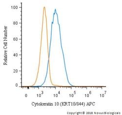
Antigen: Cytokeratin 10
Concentration: 1.0 mg/mL
Applications: Flow Cytometry, Immunohistochemistry (Paraffin), Immunofluorescence, CyTOF
Conjugate: Unconjugated
Host Species: Mouse
Research Discipline: Cytoskeleton Markers
Formulation: PBS with No Preservative
Gene ID (Entrez): 3858
Immunogen: Recombinant human KRT10 protein
Primary or Secondary: Primary
Content And Storage: Store at 4C short term. Aliquot and store at -20C long term. Avoid freeze-thaw cycles.
Molecular Weight of Antigen: 56.5 kDa
Clone: KRT10/844
Dilution: Flow Cytometry : 0.5 - 1 ug/million cells in 0.1 ml, Immunohistochemistry-Paraffin : 0.1 - 0.2 ug/ml, Immunofluorescence : 0.5 - 1.0 ug/ml, CyTOF-ready
Classification: Monoclonal
Form: Purified
Regulatory Status: RUO
Target Species: Human, Mouse
Gene Alias: BCIE, BIE, CK10, CK-10, cytokeratin 10, Cytokeratin-10, EHK, K10keratosis palmaris et plantaris, keratin 10, Keratin 10 (epidermolytic hyperkeratosis; keratosis palmaris et plantaris), keratin, type I cytoskeletal 10, keratin-10, KPP
Gene Symbols: KRT10
Isotype: IgG1 κ
Purification Method: Protein A or G purified
Test Specificity: This MAb recognizes a protein of 56.5kDa, identified as cytokeratin 10 (CK10). CK10 is expressed in all suprabasal layers of the epidermis. In the epidermis, expression of CK10 strictly parallels the extent of differentiation; it is absent in the basal layer, appears in the first suprabasal layers and increases in concentration towards the granular layer. However, CK10 is rarely detected in early stages of vulvar squamous carcinomas (tumors less than 2 cm, clinical stage I) regardless of the tumor grade. In larger and more advanced tumors (greater than 2 cm, clinical stages II and III), CK10 is detected very frequently. Expression of CK10 is related to maturation of malignant keratinocytes, being preferentially detected in more-differentiated parts.