7105846

CD53 Antibody (161-2) - Azide and BSA Free, Novus Biologicals™

Mouse Monoclonal Antibody

Manufacturer: Fischer Scientific

The price for this product is unavailable. Please request a quote

Antigen

CD53

Concentration

1.0 mg/mL

Applications

Flow Cytometry, Immunofluorescence, CyTOF

Conjugate

Unconjugated

Host Species

Mouse

Research Discipline

Immunology

Formulation

PBS with No Preservative

Gene ID (Entrez)

963

Immunogen

Stimulated human leukocytes

Primary or Secondary

Primary

Content And Storage

Store at 4C short term. Aliquot and store at -20C long term. Avoid freeze-thaw cycles.

Clone

161-2

Dilution

Flow Cytometry : 0.5 - 1 ug/million cells in 0.1 ml, Immunofluorescence : 0.5 - 1.0 ug/ml, CyTOF-ready

Classification

Monoclonal

Form

Purified

Regulatory Status

RUO

Target Species

Human, Baboon (Negative), Equine (Negative)

Gene Alias

antigen MOX44 identified by monoclonal MRC-OX44, CD53 antigentetraspanin-25, CD53 glycoprotein, CD53 molecule, CD53 tetraspan antigen, cell surface antigen, Cell surface glycoprotein CD53, MOX44transmembrane glycoprotein, Tetraspanin-25, tspan-25, TSPAN25leukocyte surface antigen CD53

Gene Symbols

CD53

Isotype

IgG2a κ

Purification Method

Protein A or G purified

Test Specificity

Recognizes a protein of 33-55kDa, identified as CD53 (Workshop VI; Code N-L033). It is expressed on monocytes and macrophages, dendritic cells, osteoblasts and osteoclasts, and on T and B cells from every stage of differentiation but is absent from platelets, red blood cells. CD53 appears to be the marker with widest reactivity as well as the marker with the strictest specificity to hematopoietic cells. CD53 is a type III membrane with both termini in the cytoplasm and two loops in the extracellular environment. This molecule, in common with other members of tetraspan family, is involved in cellular activation as part of a signal transduction complex involving other membrane glycoproteins. CD53 crosslinking induces calcium flux on human monocyte and B cells. Cross-linking of CD53 promotes activation of resting human B-lymphocytes. This MAb recognizes CD53 transfected cells and partially inhibits T-cell proliferation induced by CD3 antibody (clone: UCHT1).

Related Products

Img

Fischer Scientific

NBP247859A

--

Img

Fischer Scientific

NBP247859B

--

Img

Fischer Scientific

7105810

Mouse Monoclonal Antibody...

Img

Fischer Scientific

NBP247822A

--

Img

Fischer Scientific

NBP247822B

--

Img

Fischer Scientific

NBP232829A

--

Img

Fischer Scientific

7105364

Mouse Monoclonal Antibody has been ...

Img

Fischer Scientific

NBP232829B

--

Description

  • CD53 Monoclonal specifically detects CD53 in Human, Baboon (Negative), Equine (Negative) samples
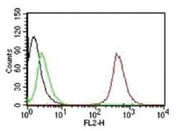
  • It is validated for Flow Cytometry, Immunocytochemistry/Immunofluorescence, Functional, Immunofluorescence, CyTOF-ready.

Compare Similar Items

Show Difference

Img

Fischer Scientific

7105846

Mouse Monoclonal Antibody...


Antigen:
CD53

Concentration:
1.0 mg/mL

Applications:
Flow Cytometry, Immunofluorescence, CyTOF

Conjugate:
Unconjugated

Host Species:
Mouse

Research Discipline:
Immunology

Formulation:
PBS with No Preservative

Gene ID (Entrez):
963

Immunogen:
Stimulated human leukocytes

Primary or Secondary:
Primary

Content And Storage:
Store at 4C short term. Aliquot and store at -20C long term. Avoid freeze-thaw cycles.

Clone:
161-2

Dilution:
Flow Cytometry : 0.5 - 1 ug/million cells in 0.1 ml, Immunofluorescence : 0.5 - 1.0 ug/ml, CyTOF-ready

Classification:
Monoclonal

Form:
Purified

Regulatory Status:
RUO

Target Species:
Human, Baboon (Negative), Equine (Negative)

Gene Alias:
antigen MOX44 identified by monoclonal MRC-OX44, CD53 antigentetraspanin-25, CD53 glycoprotein, CD53 molecule, CD53 tetraspan antigen, cell surface antigen, Cell surface glycoprotein CD53, MOX44transmembrane glycoprotein, Tetraspanin-25, tspan-25, TSPAN25leukocyte surface antigen CD53

Gene Symbols:
CD53

Isotype:
IgG2a κ

Purification Method:
Protein A or G purified

Test Specificity:
Recognizes a protein of 33-55kDa, identified as CD53 (Workshop VI; Code N-L033). It is expressed on monocytes and macrophages, dendritic cells, osteoblasts and osteoclasts, and on T and B cells from every stage of differentiation but is absent from platelets, red blood cells. CD53 appears to be the marker with widest reactivity as well as the marker with the strictest specificity to hematopoietic cells. CD53 is a type III membrane with both termini in the cytoplasm and two loops in the extracellular environment. This molecule, in common with other members of tetraspan family, is involved in cellular activation as part of a signal transduction complex involving other membrane glycoproteins. CD53 crosslinking induces calcium flux on human monocyte and B cells. Cross-linking of CD53 promotes activation of resting human B-lymphocytes. This MAb recognizes CD53 transfected cells and partially inhibits T-cell proliferation induced by CD3 antibody (clone: UCHT1).

Img

Fischer Scientific

7105848

Mouse Monoclonal Antibody...


Antigen:
Nucleolin

Concentration:
1.0 mg/mL

Applications:
Western Blot, Flow Cytometry, Immunohistochemistry (Paraffin), Immunofluorescence, CyTOF

Conjugate:
Unconjugated

Host Species:
Mouse

Research Discipline:
Cell Cycle and Replication, Chromatin Research, Core ESC Like Genes, DNA Repair, DNA replication Transcription Translation and Splicing, Stem Cell Markers

Formulation:
PBS with No Preservative

Gene ID (Entrez):
4691

Immunogen:
Lysate of SU-DHL-1 Nuclei

Primary or Secondary:
Primary

Content And Storage:
Store at 4C short term. Aliquot and store at -20C long term. Avoid freeze-thaw cycles.

Clone:
364-5

Dilution:
Western Blot : 0.5 - 1.0 ug/ml, Flow Cytometry : 0.5 - 1 ug/million cells in 0.1 ml, Immunohistochemistry-Paraffin : 0.25 - 0.5 ug/ml, Immunofluorescence : 0.5 - 1.0 ug/ml, CyTOF-ready

Classification:
Monoclonal

Form:
Purified

Regulatory Status:
RUO

Target Species:
Human, Bovine (Negative), Mouse (Negative), Rat (Negative)

Gene Alias:
C23, FLJ45706, FLJ59041, nucleolin, Protein C23

Gene Symbols:
NCL

Isotype:
IgG1 κ

Purification Method:
Protein A or G purified

Test Specificity:
Recognizes a protein of ∼76kDa, which is identified as Nucleolin (NCL). It is the major nucleolar phosphoprotein of growing eukaryotic cells. NCL is located mainly in dense fibrillar regions of the nucleolus. It is found associated with intranucleolar chromatin and pre-ribosomal particles. Human NCL gene consists of 14 exons with 13 introns and spans approximately 11kb. It induces chromatin decondensation by binding to histone H1. It is thought to play a role in pre-rRNA transcription and ribosome assembly.This MAb can be used to stain the nucleoli in cell or tissue preparations and can be used as a marker of the nucleoli in subcellular fractions. It produces a speckled pattern in the nuclei of cells of normal and malignant cells and may be used to stain the nucleoli of cells in fixed or frozen tissue sections. It can be used with paraformaldehyde fixed frozen tissue or cell preparations and formalin fixed, paraffin-embedded tissue sections.

Img

Fischer Scientific

7105850

Mouse Monoclonal Antibody...


Antigen:
VCAM-1/CD106

Concentration:
1.0 mg/mL

Applications:
Flow Cytometry, Immunofluorescence, CyTOF

Conjugate:
Unconjugated

Host Species:
Mouse

Research Discipline:
Cancer, Mesenchymal Stem Cell Markers, Stem Cell Markers

Formulation:
PBS with No Preservative

Gene ID (Entrez):
7412

Immunogen:
Activated human umbilical vein endothelial cells (HUVEC)

Primary or Secondary:
Primary

Content And Storage:
Store at 4C short term. Aliquot and store at -20C long term. Avoid freeze-thaw cycles.

Clone:
B-K9

Dilution:
Flow Cytometry : 0.5 - 1 ug/million cells in 0.1 ml, Immunofluorescence : 1 - 2 ug/ml, CyTOF-ready

Classification:
Monoclonal

Form:
Purified

Regulatory Status:
RUO

Target Species:
Human

Gene Alias:
CD106, CD106 antigen, DKFZp779G2333, INCAM-100, L1CAM, MGC99561, vascular cell adhesion molecule 1, vascular cell adhesion protein 1, V-CAM 1, VCAM1, VCAM-1

Gene Symbols:
VCAM1

Isotype:
IgG1 κ

Purification Method:
Protein A or G purified

Test Specificity:
Recognizes a protein of 110kDa, identified as CD106 (also known as vascular cell adhesion molecule-1 (VCAM-1) and INCAM-100). CD106 is a member of the Ig superfamily of adhesion molecules and is expressed at high levels on cytokine stimulated vascular endothelial cells, and at minimal levels on un-stimulated endothelial cells. It is also present on follicular and inter-follicular dendritic cells of lymph nodes, myoblasts, and some macrophages. CD106 serves as a ligand for leukocyte integrin (VLA-4 or CD49d/CD29) and mediates cell adhesion of leukocytes to activated endothelium. It plays a role in various immunological and inflammatory responses. This MAb inhibits the binding of leukocytes to VCAM-1 on stimulated endothelial cells.

Img

Fischer Scientific

7105853

Mouse Monoclonal Antibody...


Antigen:
ABO, Blood Group A Antigen

Concentration:
1.0 mg/mL

Applications:
Immunohistochemistry (Paraffin), Immunofluorescence

Conjugate:
Unconjugated

Host Species:
Mouse

Research Discipline:
__

Formulation:
PBS with No Preservative

Gene ID (Entrez):
28

Immunogen:
Mucinous colonic adenocarcinoma

Primary or Secondary:
Primary

Content And Storage:
Store at 4C short term. Aliquot and store at -20C long term. Avoid freeze-thaw cycles.

Clone:
19-OLE

Dilution:
Immunohistochemistry-Paraffin : 0.5 - 1.0 ug/ml, Immunofluorescence : 0.5 - 1.0 ug/ml

Classification:
Monoclonal

Form:
Purified

Regulatory Status:
RUO

Target Species:
Human

Gene Alias:
A transferase, A3GALNT, A3GALT1, ABO blood group (transferase A, alpha 1-3-N-acetylgalactosaminyltransferase;transferase B, alpha 1-3-galactosyltransferase), ABO glycosyltransferase, B transferase, B(A) alpha-1,3-galactosyltransferase, EC 2.4.1.37, EC 2.4.1.40, Fucosylglycoprotein 3-alpha-galactosyltransferase, Fucosylglycoprotein alpha-N-acetylgalactosaminyltransferase, Glycoprotein-fucosylgalactoside alpha-galactosyltransferase, Glycoprotein-fucosylgalactoside alpha-N-acetylgalactosaminyltransferase, GTB, Histo-blood group A transferase, histo-blood group A2 transferase, histo-blood group ABO system transferase, Histo-blood group B transferase, NAGAT

Gene Symbols:
ABO

Isotype:
IgM κ

Purification Method:
Protein A or G purified

Test Specificity:
__