Aminopeptidase N/CD13 Antibody (APN/514) - Azide and BSA Free, Novus Biologicals™
Mouse Monoclonal Antibody
Manufacturer: Fischer Scientific
The price for this product is unavailable. Please request a quote
Antigen
Aminopeptidase N/CD13
Concentration
1.0 mg/mL
Applications
Flow Cytometry, Immunohistochemistry (Paraffin), Immunofluorescence, CyTOF
Conjugate
Unconjugated
Host Species
Mouse
Research Discipline
Angiogenesis, Cellular Markers, Hematopoietic Stem Cell Markers, Immunology, Mesenchymal Stem Cell Markers, Myeloid derived Suppressor Cell, Stem Cell Markers
Formulation
PBS with No Preservative
Gene ID (Entrez)
290
Immunogen
Recombinant human CD13 protein
Primary or Secondary
Primary
Content And Storage
Store at 4C short term. Aliquot and store at -20C long term. Avoid freeze-thaw cycles.
Molecular Weight of Antigen
150 kDa
Clone
APN/514
Dilution
Flow Cytometry : 0.5 - 1 ug/million cells in 0.1 ml, Immunohistochemistry-Paraffin : 0.5 - 1.0 ug/ml, Immunofluorescence : 1 - 2 ug/ml, CyTOF-ready
Classification
Monoclonal
Form
Purified
Regulatory Status
RUO
Target Species
Human
Gene Alias
alanyl (membrane) aminopeptidase, Alanyl aminopeptidase, Aminopeptidase M, aminopeptidase N, AP-M, AP-N, CD13 antigen, CD13APN, EC 3.4.11, EC 3.4.11.2, gp150, LAP1, Microsomal aminopeptidase, Myeloid plasma membrane glycoprotein CD13, P150, PEPNhAPN
Gene Symbols
ANPEP
Isotype
IgG1 κ
Purification Method
Protein A or G purified
Test Specificity
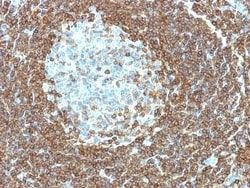
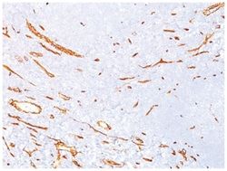
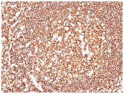
This MAb recognizes an extracellular epitope of an integral membrane glycoprotein of 150kDa, identified as CD13. This antigen is present on most cells of myeloid origin including granulocytes, monocytes, mast cells, and GM-progenitor cells. It is also expressed by the majority of AML, CML in myeloid blast crisis, and in a smaller fraction of lymphoid leukemias. It is absent from normal lymphocytes, platelets and erythrocytes. CD13 is also present on fibroblasts; endothelial cells, epithelial cells from renal proximal tubules and intestinal brush border, bone marrow stromal cells, osteoclasts, and cells lining bile duct canaliculi. CD13 is identical to aminopeptidase N (APN), a prominent membrane-bound metalloprotease present on the surface of intestinal brush border and renal tubules. CD13 plays a role in metabolism of biologically active peptides, in phagocytosis, and in bactericidal/tumoricidal activities. It also serves as a receptor for human coronaviruses (HCV). The lineage-restricted pattern of expression of CD13 within the hemopoietic compartment suggests that it may be important in myeloid cell differentiation.
Related Products

Description
- Aminopeptidase N/CD13 Monoclonal specifically detects Aminopeptidase N/CD13 in Human samples
- It is validated for ELISA.
Compare Similar Items
Show Difference
Antigen: Aminopeptidase N/CD13
Concentration: 1.0 mg/mL
Applications: Flow Cytometry, Immunohistochemistry (Paraffin), Immunofluorescence, CyTOF
Conjugate: Unconjugated
Host Species: Mouse
Research Discipline: Angiogenesis, Cellular Markers, Hematopoietic Stem Cell Markers, Immunology, Mesenchymal Stem Cell Markers, Myeloid derived Suppressor Cell, Stem Cell Markers
Formulation: PBS with No Preservative
Gene ID (Entrez): 290
Immunogen: Recombinant human CD13 protein
Primary or Secondary: Primary
Content And Storage: Store at 4C short term. Aliquot and store at -20C long term. Avoid freeze-thaw cycles.
Molecular Weight of Antigen: 150 kDa
Clone: APN/514
Dilution: Flow Cytometry : 0.5 - 1 ug/million cells in 0.1 ml, Immunohistochemistry-Paraffin : 0.5 - 1.0 ug/ml, Immunofluorescence : 1 - 2 ug/ml, CyTOF-ready
Classification: Monoclonal
Form: Purified
Regulatory Status: RUO
Target Species: Human
Gene Alias: alanyl (membrane) aminopeptidase, Alanyl aminopeptidase, Aminopeptidase M, aminopeptidase N, AP-M, AP-N, CD13 antigen, CD13APN, EC 3.4.11, EC 3.4.11.2, gp150, LAP1, Microsomal aminopeptidase, Myeloid plasma membrane glycoprotein CD13, P150, PEPNhAPN
Gene Symbols: ANPEP
Isotype: IgG1 κ
Purification Method: Protein A or G purified
Test Specificity: This MAb recognizes an extracellular epitope of an integral membrane glycoprotein of 150kDa, identified as CD13. This antigen is present on most cells of myeloid origin including granulocytes, monocytes, mast cells, and GM-progenitor cells. It is also expressed by the majority of AML, CML in myeloid blast crisis, and in a smaller fraction of lymphoid leukemias. It is absent from normal lymphocytes, platelets and erythrocytes. CD13 is also present on fibroblasts; endothelial cells, epithelial cells from renal proximal tubules and intestinal brush border, bone marrow stromal cells, osteoclasts, and cells lining bile duct canaliculi. CD13 is identical to aminopeptidase N (APN), a prominent membrane-bound metalloprotease present on the surface of intestinal brush border and renal tubules. CD13 plays a role in metabolism of biologically active peptides, in phagocytosis, and in bactericidal/tumoricidal activities. It also serves as a receptor for human coronaviruses (HCV). The lineage-restricted pattern of expression of CD13 within the hemopoietic compartment suggests that it may be important in myeloid cell differentiation.
Antigen: Myosin heavy chain 11
Concentration: 1.0 mg/mL
Applications: Flow Cytometry, Immunohistochemistry (Paraffin), Immunofluorescence, CyTOF
Conjugate: Unconjugated
Host Species: Mouse
Research Discipline: Hypoxia, Stem Cell Markers
Formulation: PBS with No Preservative
Gene ID (Entrez): 4629
Immunogen: Human uterus extract
Primary or Secondary: Primary
Content And Storage: Store at 4C short term. Aliquot and store at -20C long term. Avoid freeze-thaw cycles.
Molecular Weight of Antigen: __
Clone: ID8
Dilution: Flow Cytometry : 0.5 - 1 ug/million cells in 0.1 ml, Immunohistochemistry-Paraffin : 0.5 - 1.0 ug/ml, Immunofluorescence : 0.5 - 1.0 ug/ml, CyTOF-ready
Classification: Monoclonal
Form: Purified
Regulatory Status: RUO
Target Species: Human, Rat, Porcine, Canine, Chicken, Feline, Guinea Pig, Primate, Rabbit
Gene Alias: AAT4, FAA4, MYH11, MYH11 myosin, heavy chain 11, smooth muscle, SMHC, SMMHC, Smooth muscle myosin heavy chain
Gene Symbols: MYH11
Isotype: IgG1 κ
Purification Method: Protein A or G purified
Test Specificity: Smooth muscle myosin heavy chain (SM-MHC) is a cytoplasmic structural protein, which is a major component of the contractile apparatus in smooth muscle cells. Expression of smooth muscle myosin is developmentally regulated, appearing early in smooth muscle development, and is specific for smooth muscle development. Two isoforms of smooth muscle myosin heavy chain have been identified, designated MHC-1 and MHC-2. The antibody may be useful for the study of breast tumors as the presence of an intact layer of myoepithelial cells is an important feature, which may distinguish benign breast lesions and carcinoma in situ from invasive tumors.
Antigen: Myosin heavy chain 11
Concentration: 1.0 mg/mL
Applications: Flow Cytometry, Immunohistochemistry (Paraffin), Immunofluorescence, CyTOF
Conjugate: Unconjugated
Host Species: Mouse
Research Discipline: Hypoxia, Stem Cell Markers
Formulation: PBS with No Preservative
Gene ID (Entrez): 4629
Immunogen: Recombinant human MYH11 protein
Primary or Secondary: Primary
Content And Storage: Store at 4C short term. Aliquot and store at -20C long term. Avoid freeze-thaw cycles.
Molecular Weight of Antigen: __
Clone: MYH11/923
Dilution: Flow Cytometry : 0.5 - 1 ug/million cells in 0.1 ml, Immunohistochemistry-Paraffin : 0.5 - 1.0 ug/ml, Immunofluorescence : 0.5 - 1.0 ug/ml, CyTOF-ready
Classification: Monoclonal
Form: Purified
Regulatory Status: RUO
Target Species: Human, Rat
Gene Alias: AAT4, FAA4, MYH11, MYH11 myosin, heavy chain 11, smooth muscle, SMHC, SMMHC, Smooth muscle myosin heavy chain
Gene Symbols: MYH11
Isotype: IgG1 κ
Purification Method: Protein A or G purified
Test Specificity: Smooth muscle myosin heavy chain (SM-MHC) is a cytoplasmic structural protein, which is a major component of the contractile apparatus in smooth muscle cells. Expression of smooth muscle myosin is developmentally regulated, appearing early in smooth muscle development, and is specific for smooth muscle development. Two isoforms of smooth muscle myosin heavy chain have been identified, designated MHC-1 and MHC-2. The antibody may be useful for the study of breast tumors as the presence of an intact layer of myoepithelial cells is an important feature, which may distinguish benign breast lesions and carcinoma in situ from invasive tumors.