Pax7 Antibody (PAX7/1187) - Azide and BSA Free, Novus Biologicals™
Mouse Monoclonal Antibody
Manufacturer: Fischer Scientific
The price for this product is unavailable. Please request a quote
Antigen
Pax7
Concentration
1.0 mg/mL
Applications
Immunohistochemistry (Paraffin)
Conjugate
Unconjugated
Host Species
Mouse
Research Discipline
Apoptosis, Mesenchymal Stem Cell Markers, Stem Cell Markers
Formulation
PBS with No Preservative
Gene ID (Entrez)
5081
Immunogen
Recombinant fragment (aa300-600) of human PAX7 protein
Primary or Secondary
Primary
Content And Storage
Store at 4C short term. Aliquot and store at -20C long term. Avoid freeze-thaw cycles.
Molecular Weight of Antigen
57 kDa
Clone
PAX7/1187
Dilution
Immunohistochemistry-Paraffin : 0.5 - 1.0 ug/ml
Classification
Monoclonal
Form
Purified
Regulatory Status
RUO
Target Species
Human
Gene Alias
FLJ37460, HuP1, paired box 7, paired box gene 7, paired box homeotic gene 7, paired box protein Pax-7, paired domain gene 7, PAX7 transcriptional factor, PAX7B, RMS2
Gene Symbols
PAX7
Isotype
IgG1 κ
Purification Method
Protein A or G purified
Test Specificity
The Pax gene family of nuclear transcription factors is comprised of nine members that function during embryogenesis to regulate the temporal and position-dependent differentiation of cells. In addition, the family is involved in a variety of signal transduction pathways in the adult organism. Mutations in the Pax family of proteins have been linked to disease and cancer in humans. Pax-7 is a protein specifically expressed in cultured satellite cell-derived myoblasts. In situ hybridization reveals that Pax-7 is also expressed in satellite cells residing in adult muscle. A chromosomal aberration in the gene encoding Pax-7 causes rhabdomyosarcoma 2 (RMS2) (also called alveolar rhabdomyosarcoma).
Related Products


Description
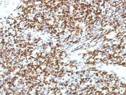
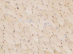
- Pax7 Monoclonal specifically detects Pax7 in Human samples
- It is validated for Immunohistochemistry, Immunohistochemistry-Paraffin.
Compare Similar Items
Show Difference
Antigen: Pax7
Concentration: 1.0 mg/mL
Applications: Immunohistochemistry (Paraffin)
Conjugate: Unconjugated
Host Species: Mouse
Research Discipline: Apoptosis, Mesenchymal Stem Cell Markers, Stem Cell Markers
Formulation: PBS with No Preservative
Gene ID (Entrez): 5081
Immunogen: Recombinant fragment (aa300-600) of human PAX7 protein
Primary or Secondary: Primary
Content And Storage: Store at 4C short term. Aliquot and store at -20C long term. Avoid freeze-thaw cycles.
Molecular Weight of Antigen: 57 kDa
Clone: PAX7/1187
Dilution: Immunohistochemistry-Paraffin : 0.5 - 1.0 ug/ml
Classification: Monoclonal
Form: Purified
Regulatory Status: RUO
Target Species: Human
Gene Alias: FLJ37460, HuP1, paired box 7, paired box gene 7, paired box homeotic gene 7, paired box protein Pax-7, paired domain gene 7, PAX7 transcriptional factor, PAX7B, RMS2
Gene Symbols: PAX7
Isotype: IgG1 κ
Purification Method: Protein A or G purified
Test Specificity: The Pax gene family of nuclear transcription factors is comprised of nine members that function during embryogenesis to regulate the temporal and position-dependent differentiation of cells. In addition, the family is involved in a variety of signal transduction pathways in the adult organism. Mutations in the Pax family of proteins have been linked to disease and cancer in humans. Pax-7 is a protein specifically expressed in cultured satellite cell-derived myoblasts. In situ hybridization reveals that Pax-7 is also expressed in satellite cells residing in adult muscle. A chromosomal aberration in the gene encoding Pax-7 causes rhabdomyosarcoma 2 (RMS2) (also called alveolar rhabdomyosarcoma).
Antigen: Pax7
Concentration: 1.0 mg/mL
Applications: Immunohistochemistry (Paraffin)
Conjugate: Unconjugated
Host Species: Mouse
Research Discipline: Apoptosis, Mesenchymal Stem Cell Markers, Stem Cell Markers
Formulation: PBS with No Preservative
Gene ID (Entrez): 5081
Immunogen: Recombinant fragment (aa300-600) of human PAX7 protein
Primary or Secondary: Primary
Content And Storage: Store at 4C short term. Aliquot and store at -20C long term. Avoid freeze-thaw cycles.
Molecular Weight of Antigen: 57 kDa
Clone: SPM613
Dilution: Immunohistochemistry-Paraffin : 0.5 - 1.0 ug/ml
Classification: Monoclonal
Form: Purified
Regulatory Status: RUO
Target Species: Human
Gene Alias: FLJ37460, HuP1, paired box 7, paired box gene 7, paired box homeotic gene 7, paired box protein Pax-7, paired domain gene 7, PAX7 transcriptional factor, PAX7B, RMS2
Gene Symbols: PAX7
Isotype: IgG1 κ
Purification Method: Protein A or G purified
Test Specificity: The Pax gene family of nuclear transcription factors is comprised of nine members that function during embryogenesis to regulate the temporal and position-dependent differentiation of cells. In addition, the family is involved in a variety of signal transduction pathways in the adult organism. Mutations in the Pax family of proteins have been linked to disease and cancer in humans. Pax-7 is a protein specifically expressed in cultured satellite cell-derived myoblasts. In situ hybridization reveals that Pax-7 is also expressed in satellite cells residing in adult muscle. A chromosomal aberration in the gene encoding Pax-7 causes rhabdomyosarcoma 2 (RMS2) (also called alveolar rhabdomyosarcoma).
Antigen: Blood Group Lewis b
Concentration: 1.0 mg/mL
Applications: Immunohistochemistry (Paraffin), Immunofluorescence
Conjugate: Unconjugated
Host Species: Mouse
Research Discipline: __
Formulation: PBS with No Preservative
Gene ID (Entrez): 2525
Immunogen: Mucin isolated from a human ovarian cyst fluid
Primary or Secondary: Primary
Content And Storage: Store at 4C short term. Aliquot and store at -20C long term. Avoid freeze-thaw cycles.
Molecular Weight of Antigen: 45 kDa
Clone: SPM194
Dilution: Immunohistochemistry-Paraffin : 0.5 - 1.0 ug/ml, Immunofluorescence : 0.5 - 1.0 ug/ml
Classification: Monoclonal
Form: Purified
Regulatory Status: RUO
Target Species: Human, Guinea Pig
Gene Alias: alpha-(1,3/1,4)-fucosyltransferase, Blood group Lewis alpha-4-fucosyltransferase, CD174, EC 2.4.1, EC 2.4.1.65, FT3B, Fucosyltransferase 3, fucosyltransferase 3 (galactoside 3(4)-L-fucosyltransferase, Lewis blood group), Fucosyltransferase III, FucT-III, galactoside 3(4)-L-fucosyltransferase, LEfucosyltransferase 3 (galactoside 3(4)-L-fucosyltransferase, Lewis blood groupincluded), Les, Lewis FT, MGC131739
Gene Symbols: FUT3
Isotype: IgG1 κ
Purification Method: Protein A or G purified
Test Specificity: The Lewis histo-blood group system comprises a set of fucosylated glycosphingolipids that are synthesized by exocrine epithelial cells and circulate in body fluids. The glycosphingolipids function in embryogenesis, tissue differentiation, tumor metastasis, inflammation, and bacterial adhesion. They are secondarily absorbed to red blood cells giving rise to their Lewis phenotype. This gene is a member of the fucosyltransferase family, which catalyzes the addition of fucose to precursor polysaccharides in the last step of Lewis antigen biosynthesis. It encodes an enzyme with alpha(1,3)-fucosyltransferase and alpha(1,4)-fucosyltransferase activities. Lewis blood group antigens are carbohydrate moieties structurally integrated in mucous secretions. Lewis antigen system alterations have been described in gastric carcinoma and associated lesions. Anomalous expression of Lewis B antigen has been found in some non-secretory gastric carcinomas and colorectal cancers.