Enolase 2/Neuron-specific Enolase Mouse anti-Human, Clone: ENO2/1462, Novus Biologicals™
Mouse Monoclonal Antibody
Manufacturer: Fischer Scientific
The price for this product is unavailable. Please request a quote
Antigen
Enolase 2/Neuron-specific Enolase
Concentration
0.2 mg/mL
Applications
Flow Cytometry, Immunocytochemistry, Immunofluorescence, Immunohistochemistry (Paraffin), SDS-Page
Conjugate
Unconjugated
Host Species
Mouse
Research Discipline
Cancer, Cellular Markers, Neuronal Cell Markers, Neuroscience
Formulation
10mM PBS with 0.05% BSA with 0.05% Sodium Azide
Gene ID (Entrez)
2026
Immunogen
A synthetic peptide of human NSE gamma (around aa416-433; exact sequence is proprietary)
Primary or Secondary
Primary
Content And Storage
Store at 4C short term. Aliquot and store at -20C long term. Avoid freeze-thaw cycles.
Molecular Weight of Antigen
50 kDa
Clone
ENO2/1462
Dilution
Flow Cytometry 0.5-1 ug/million cells, Immunocytochemistry/Immunofluorescence 1-2 ug/ml, Immunohistochemistry-Paraffin 0.1-0.2 ug/ml, SDS-Page, Protein Array 1:100-1:2000
Classification
Monoclonal
Form
Purified
Regulatory Status
RUO
Target Species
Human
Gene Alias
2-phospho-D-glycerate hydrolyase, 2-phospho-D-glycerate hydro-lyase, EC 4.2.1.11, Enolase 2, enolase 2 (gamma, neuronal), gamma-enolase, Neural enolase, neuron specific gamma enolase, neurone-specific enolase, Neuron-specific enolase, NSE
Gene Symbols
ENO2
Isotype
IgG2b
Purification Method
Protein A or G purified
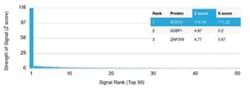
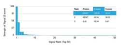
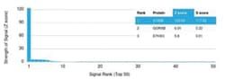
Test Specificity
The specificity of this monoclonal antibody to its intended target was validated by HuProtTM Array, containing more than 19,000, full-length human proteins. Recognizes a protein of about 50kDa, which is identified as gamma -enolase. Three isoenzymes of enolases are identified, alpha, beta and gamma. Alpha-isoform is expressed in most tissues, whereas beta-form is expressed predominantly in muscle tissue whereas gamma-enolase is found only in nervous tissue. These isoforms exist as both homodimers and heterodimers, and they play a role in converting phosphoglyceric acid to phosphenolpyruvic acid in the glycolytic pathway. NSE-gamma is a useful marker to identify peripheral nerves and tumors of neuro-endocrine origins, such as pheochromocytomas. It it be usually employed in combination with other markers such as Synaptophysin, Chromogranin A, and Neurofilament.
Related Products

Description
- Enolase 2/Neuron-specific Enolase Monoclonal specifically detects Enolase 2/Neuron-specific Enolase in Human samples
- It is validated for Immunohistochemistry, Immunohistochemistry-Paraffin, Protein Array.
Compare Similar Items
Show Difference
Antigen: Enolase 2/Neuron-specific Enolase
Concentration: 0.2 mg/mL
Applications: Flow Cytometry, Immunocytochemistry, Immunofluorescence, Immunohistochemistry (Paraffin), SDS-Page
Conjugate: Unconjugated
Host Species: Mouse
Research Discipline: Cancer, Cellular Markers, Neuronal Cell Markers, Neuroscience
Formulation: 10mM PBS with 0.05% BSA with 0.05% Sodium Azide
Gene ID (Entrez): 2026
Immunogen: A synthetic peptide of human NSE gamma (around aa416-433; exact sequence is proprietary)
Primary or Secondary: Primary
Content And Storage: Store at 4C short term. Aliquot and store at -20C long term. Avoid freeze-thaw cycles.
Molecular Weight of Antigen: 50 kDa
Clone: ENO2/1462
Dilution: Flow Cytometry 0.5-1 ug/million cells, Immunocytochemistry/Immunofluorescence 1-2 ug/ml, Immunohistochemistry-Paraffin 0.1-0.2 ug/ml, SDS-Page, Protein Array 1:100-1:2000
Classification: Monoclonal
Form: Purified
Regulatory Status: RUO
Target Species: Human
Gene Alias: 2-phospho-D-glycerate hydrolyase, 2-phospho-D-glycerate hydro-lyase, EC 4.2.1.11, Enolase 2, enolase 2 (gamma, neuronal), gamma-enolase, Neural enolase, neuron specific gamma enolase, neurone-specific enolase, Neuron-specific enolase, NSE
Gene Symbols: ENO2
Isotype: IgG2b
Purification Method: Protein A or G purified
Test Specificity: The specificity of this monoclonal antibody to its intended target was validated by HuProtTM Array, containing more than 19,000, full-length human proteins. Recognizes a protein of about 50kDa, which is identified as gamma -enolase. Three isoenzymes of enolases are identified, alpha, beta and gamma. Alpha-isoform is expressed in most tissues, whereas beta-form is expressed predominantly in muscle tissue whereas gamma-enolase is found only in nervous tissue. These isoforms exist as both homodimers and heterodimers, and they play a role in converting phosphoglyceric acid to phosphenolpyruvic acid in the glycolytic pathway. NSE-gamma is a useful marker to identify peripheral nerves and tumors of neuro-endocrine origins, such as pheochromocytomas. It it be usually employed in combination with other markers such as Synaptophysin, Chromogranin A, and Neurofilament.
Antigen: Factor XIIIa
Concentration: 0.2 mg/mL
Applications: Western Blot, Flow Cytometry, ELISA, Immunocytochemistry, Immunofluorescence, Immunohistochemistry (Paraffin)
Conjugate: Unconjugated
Host Species: Mouse
Research Discipline: Apoptosis, Cancer, Cell Biology
Formulation: 10mM PBS with 0.05% BSA with 0.05% Sodium Azide
Gene ID (Entrez): 2162
Immunogen: Recombinant fragment of human Factor XIIIa protein (aa46-181) (exact sequence is proprietary)
Primary or Secondary: Primary
Content And Storage: Store at 4C short term. Aliquot and store at -20C long term. Avoid freeze-thaw cycles.
Molecular Weight of Antigen: 83 kDa
Clone: F13A1/1448
Dilution: Western Blot 0.5-1.0 ug/ml, Flow Cytometry 0.5-1 ug/million cells, ELISA 2-4 ug/ml, Immunocytochemistry/Immunofluorescence 0.5-1 ug/ml, Immunohistochemistry-Paraffin 1-2 ug/ml, SDS-Page, Protein Array 1:100-1:2000
Classification: Monoclonal
Form: Purified
Regulatory Status: RUO
Target Species: Human
Gene Alias: bA525O21.1 (coagulation factor XIII, A1 polypeptide), coagulation factor XIII A chain, coagulation factor XIII, A1 polypeptide, Coagulation factor XIIIa, EC 2.3.2.13, F13Acoagulation factor XIII, A polypeptide, factor XIIIa, fibrin stabilizing factor, A subunit, fibrinoligase, FSF, A subunit, Protein-glutamine gamma-glutamyltransferase A chain, TGase, Transglutaminase A chain, transglutaminase. plasma
Gene Symbols: F13A1
Isotype: IgG2b κ
Purification Method: Protein A or G purified
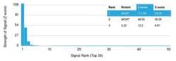
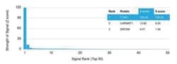
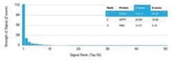
Test Specificity: The specificity of this monoclonal antibody to its intended target was validated by HuProtTM Array, containing more than 19,000, full-length human proteins. It recognizes a protein of 83kDa, which is identified as Factor XIIIa. It has been identified in platelets, megakaryocytes, and fibroblast-like mesenchymal or histiocytic cells in the placenta, uterus, and prostate, monocytes and macrophages and dermal dendritic cells. Anti-factor XIIIa has been found to be useful in differentiating between dermatofibroma (almost all cases are positive), dermatofibrosarcoma protuberans ( -/+) and desmoplastic malignant melanoma (-). Anti-factor XIIIa positivity is also seen in capillary hemagioblastoma, hemangioendothelioma, hemangiopericytoma, xanthogranuloma, xanthoma, hepatocellular carcinoma, glomus tumor, and meningioma.
Antigen: Factor XIIIa
Concentration: 0.2 mg/mL
Applications: Western Blot, Flow Cytometry, ELISA, Immunocytochemistry, Immunofluorescence, Immunohistochemistry (Paraffin)
Conjugate: Unconjugated
Host Species: Mouse
Research Discipline: Apoptosis, Cancer, Cell Biology
Formulation: 10mM PBS with 0.05% BSA with 0.05% Sodium Azide
Gene ID (Entrez): 2162
Immunogen: Recombinant fragment of human Factor XIIIa protein (aa46-181) (exact sequence is proprietary)
Primary or Secondary: Primary
Content And Storage: Store at 4C short term. Aliquot and store at -20C long term. Avoid freeze-thaw cycles.
Molecular Weight of Antigen: 83 kDa
Clone: F13A1/1448
Dilution: Western Blot 0.5-1.0 ug/ml, Flow Cytometry 0.5-1 ug/million cells, ELISA 2-4 ug/ml, Immunocytochemistry/Immunofluorescence 0.5-1 ug/ml, Immunohistochemistry-Paraffin 1-2 ug/ml, SDS-Page, Protein Array 1:100-1:2000
Classification: Monoclonal
Form: Purified
Regulatory Status: RUO
Target Species: Human
Gene Alias: bA525O21.1 (coagulation factor XIII, A1 polypeptide), coagulation factor XIII A chain, coagulation factor XIII, A1 polypeptide, Coagulation factor XIIIa, EC 2.3.2.13, F13Acoagulation factor XIII, A polypeptide, factor XIIIa, fibrin stabilizing factor, A subunit, fibrinoligase, FSF, A subunit, Protein-glutamine gamma-glutamyltransferase A chain, TGase, Transglutaminase A chain, transglutaminase. plasma
Gene Symbols: F13A1
Isotype: IgG2b κ
Purification Method: Protein A or G purified
Test Specificity: The specificity of this monoclonal antibody to its intended target was validated by HuProtTM Array, containing more than 19,000, full-length human proteins. It recognizes a protein of 83kDa, which is identified as Factor XIIIa. It has been identified in platelets, megakaryocytes, and fibroblast-like mesenchymal or histiocytic cells in the placenta, uterus, and prostate, monocytes and macrophages and dermal dendritic cells. Anti-factor XIIIa has been found to be useful in differentiating between dermatofibroma (almost all cases are positive), dermatofibrosarcoma protuberans ( -/+) and desmoplastic malignant melanoma (-). Anti-factor XIIIa positivity is also seen in capillary hemagioblastoma, hemangioendothelioma, hemangiopericytoma, xanthogranuloma, xanthoma, hepatocellular carcinoma, glomus tumor, and meningioma.