MCM7 Mouse anti-Human, Clone: MCM7/1467, Novus Biologicals™
Mouse Monoclonal Antibody
Manufacturer: Fischer Scientific
The price for this product is unavailable. Please request a quote
Antigen
MCM7
Concentration
0.2 mg/mL
Applications
Flow Cytometry, Immunocytochemistry, Immunofluorescence, Immunohistochemistry (Paraffin), SDS-Page
Conjugate
Unconjugated
Host Species
Mouse
Research Discipline
Cell Cycle and Replication, Cellular Markers, Core ESC Like Genes, DNA Repair, Stem Cell Markers
Formulation
0mM PBS with 0.05% BSA with 0.05% Sodium Azide
Gene ID (Entrez)
4176
Immunogen
Recombinant human MCM7 protein fragment (aa195-319) (exact sequence is proprietary)
Primary or Secondary
Primary
Content And Storage
Store at 4C short term. Aliquot and store at -20C long term. Avoid freeze-thaw cycles.
Molecular Weight of Antigen
88 kDa
Clone
MCM7/1467
Dilution
Flow Cytometry 0.5-1 ug/million cells, Immunocytochemistry/Immunofluorescence 0.5-1 ug/ml, Immunohistochemistry-Paraffin 1-2 ug/ml, SDS-Page, Protein Array 1:100-1:2000
Classification
Monoclonal
Form
Purified
Regulatory Status
RUO
Target Species
Human
Gene Alias
CDC47 homolog, CDC47P85MCM, DNA replication licensing factor MCM7, EC 3.6.4.12, homolog of S. cerevisiae Cdc47, MCM2PNAS146, MCM7 minichromosome maintenance deficient 7 (S. cerevisiae), minichromosome maintenance complex component 7, minichromosome maintenance deficient (S. cerevisiae) 7, minichromosome maintenance deficient 7, P1.1-MCM3, P1CDC47
Gene Symbols
MCM7
Isotype
IgG2a κ
Purification Method
Protein A or G purified
Test Specificity
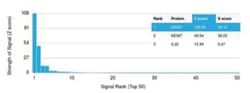
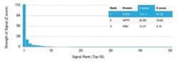
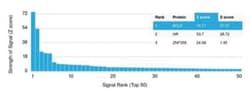
The specificity of this monoclonal antibody to its intended target was validated by HuProt Array, containing more than 19,000, full-length human proteins. MCM7 is one of the highly conserved mini-chromosome maintenance proteins (MCM) that is essential for the initiation of eukaryotic genome replication. The hexameric protein complex formed by theMCM proteins is a key component of the pre-replication complex and may be involved in the formation of replication forks and in the recruitment of other DNA replication related proteins. The MCM complex consisting of this protein and MCM2, 4 and 6 proteins possesses DNA helicase activity, and may act as a DNA unwinding enzyme. Cyclin D1-dependent kinase, CDK4, is found to associate with this protein, and may regulate the binding of this protein with the tumor suppressor protein RB1/RB.
Related Products

Description
- Description MCM7 Monoclonal specifically detects MCM7 in Human samples
- It is validated for Western Blot, Flow Cytometry, Immunohistochemistry, Immunocytochemistry/Immunofluorescence, Immunohistochemistry-Paraffin, Protein Array.
Compare Similar Items
Show Difference
Antigen: MCM7
Concentration: 0.2 mg/mL
Applications: Flow Cytometry, Immunocytochemistry, Immunofluorescence, Immunohistochemistry (Paraffin), SDS-Page
Conjugate: Unconjugated
Host Species: Mouse
Research Discipline: Cell Cycle and Replication, Cellular Markers, Core ESC Like Genes, DNA Repair, Stem Cell Markers
Formulation: 0mM PBS with 0.05% BSA with 0.05% Sodium Azide
Gene ID (Entrez): 4176
Immunogen: Recombinant human MCM7 protein fragment (aa195-319) (exact sequence is proprietary)
Primary or Secondary: Primary
Content And Storage: Store at 4C short term. Aliquot and store at -20C long term. Avoid freeze-thaw cycles.
Molecular Weight of Antigen: 88 kDa
Clone: MCM7/1467
Dilution: Flow Cytometry 0.5-1 ug/million cells, Immunocytochemistry/Immunofluorescence 0.5-1 ug/ml, Immunohistochemistry-Paraffin 1-2 ug/ml, SDS-Page, Protein Array 1:100-1:2000
Classification: Monoclonal
Form: Purified
Regulatory Status: RUO
Target Species: Human
Gene Alias: CDC47 homolog, CDC47P85MCM, DNA replication licensing factor MCM7, EC 3.6.4.12, homolog of S. cerevisiae Cdc47, MCM2PNAS146, MCM7 minichromosome maintenance deficient 7 (S. cerevisiae), minichromosome maintenance complex component 7, minichromosome maintenance deficient (S. cerevisiae) 7, minichromosome maintenance deficient 7, P1.1-MCM3, P1CDC47
Gene Symbols: MCM7
Isotype: IgG2a κ
Purification Method: Protein A or G purified
Test Specificity: The specificity of this monoclonal antibody to its intended target was validated by HuProt Array, containing more than 19,000, full-length human proteins. MCM7 is one of the highly conserved mini-chromosome maintenance proteins (MCM) that is essential for the initiation of eukaryotic genome replication. The hexameric protein complex formed by theMCM proteins is a key component of the pre-replication complex and may be involved in the formation of replication forks and in the recruitment of other DNA replication related proteins. The MCM complex consisting of this protein and MCM2, 4 and 6 proteins possesses DNA helicase activity, and may act as a DNA unwinding enzyme. Cyclin D1-dependent kinase, CDK4, is found to associate with this protein, and may regulate the binding of this protein with the tumor suppressor protein RB1/RB.
Antigen: MCM7
Concentration: 0.2 mg/mL
Applications: __
Conjugate: Unconjugated
Host Species: Mouse
Research Discipline: Cell Cycle and Replication, Cellular Markers, Core ESC Like Genes, DNA Repair, Stem Cell Markers
Formulation: 10mM PBS with 0.05% BSA with 0.05% Sodium Azide
Gene ID (Entrez): 4176
Immunogen: Recombinant human MCM7 protein fragment (aa195-319) (exact sequence is proprietary)
Primary or Secondary: Primary
Content And Storage: Store at 4C short term. Aliquot and store at -20C long term. Avoid freeze-thaw cycles.
Molecular Weight of Antigen: 88 kDa
Clone: MCM7/1469
Dilution: Flow Cytometry 0.5-1 ug/million cells, Immunocytochemistry/Immunofluorescence 0.5-1 ug/ml, Immunohistochemistry-Paraffin 0.5-1 ug/ml, SDS-Page, Protein Array 1:100-1:2000
Classification: Monoclonal
Form: Purified
Regulatory Status: RUO
Target Species: Human
Gene Alias: CDC47 homolog, CDC47P85MCM, DNA replication licensing factor MCM7, EC 3.6.4.12, homolog of S. cerevisiae Cdc47, MCM2PNAS146, MCM7 minichromosome maintenance deficient 7 (S. cerevisiae), minichromosome maintenance complex component 7, minichromosome maintenance deficient (S. cerevisiae) 7, minichromosome maintenance deficient 7, P1.1-MCM3, P1CDC47
Gene Symbols: MCM7
Isotype: IgG2b κ
Purification Method: Protein A or G purified
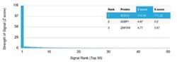
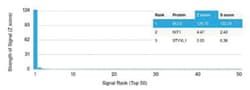
Test Specificity: The specificity of this monoclonal antibody to its intended target was validated by HuProtTM Array, containing more than 19,000, full-length human proteins. MCM7 is one of the highly conserved mini-chromosome maintenance proteins (MCM) that is essential for the initiation of eukaryotic genome replication. The hexameric protein complex formed by theMCM proteins is a key component of the pre-replication complex and may be involved in the formation of replication forks and in the recruitment of other DNA replication related proteins. The MCM complex consisting of this protein and MCM2, 4 and 6 proteins possesses DNA helicase activity, and may act as a DNA unwinding enzyme. Cyclin D1-dependent kinase, CDK4, is found to associate with this protein, and may regulate the binding of this protein with the tumor suppressor protein RB1/RB.
Antigen: S100B
Concentration: 0.2 mg/mL
Applications: Western Blot, Flow Cytometry, Immunocytochemistry, Immunofluorescence, Immunohistochemistry (Paraffin), SDS-Page
Conjugate: Unconjugated
Host Species: Mouse
Research Discipline: Apoptosis, Cytoskeleton Markers, Neuroscience, Stem Cells
Formulation: 10mM PBS with 0.05% BSA with 0.05% Sodium Azide
Gene ID (Entrez): 6285
Immunogen: Recombinant full-length human S100B protein
Primary or Secondary: Primary
Content And Storage: Store at 4C short term. Aliquot and store at -20C long term. Avoid freeze-thaw cycles.
Molecular Weight of Antigen: 13 kDa
Clone: S100B/1012
Dilution: Western Blot 0.5-1 ug/ml, Flow Cytometry 0.5-1 ug/million cells, Immunocytochemistry/Immunofluorescence 1-2 ug/ml, Immunohistochemistry-Paraffin 0.25-0.5 ug/ml, SDS-Page, Protein Array 1:100-1:2000
Classification: Monoclonal
Form: Purified
Regulatory Status: RUO
Target Species: Human, Mouse, Rat, Bovine
Gene Alias: beta (neural), NEF, S100, S100 beta, S100 calcium binding protein B, S100 calcium-binding protein B, S100 calcium-binding protein, beta (neural), S-100 calcium-binding protein, beta chain, 10protein S100-B, S-100 protein beta chain, S-100 protein subunit beta, S100beta
Gene Symbols: S100B
Isotype: IgG2a κ
Purification Method: Protein A or G purified
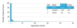
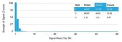
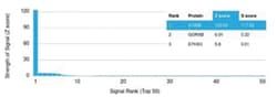
Test Specificity: The specificity of this monoclonal antibody to its intended target was validated by HuProtTM Array, containing more than 19,000, full-length human proteins. S100 belongs to the family of calcium binding proteins. S100A and S100B proteins are two members of the S100 family. S100A is composed of an alpha and a beta chain whereas S100B is composed of two beta chains. This antibody is specific against an epitope located on the beta -chain (i.e. in S-100A and S-100B) but not on the alpha-chain of S-100 (i.e. in S-100A and S100A0). This antibody can be used to localize S-100A and S-100B in various tissue sections. S-100 protein has been found in normal melanocytes, Langerhans cells, histiocytes, chondrocytes, lip ocytes, skeletal and cardiac muscle, Schwann cells, epithelial and myoepithelial cells of the breast, salivary and sweat glands, as well as in glial cells. Neoplasms derived from these cells also express S-100 protein, albeit non-uniformly. A large number of well-differentiated tumo