CD34, Mouse anti-Monkey, Human, Clone: QBEnd/10, Millipore Sigma™
Mouse Monoclonal Antibody
Manufacturer: Fischer Scientific
The price for this product is unavailable. Please request a quote
Antigen
CD34
Dilution
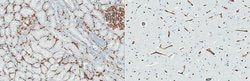
Immunohistochemistry (Paraffin) Analysis: A 1:250 dilution from a representative lot detected CD34 in human brain tissue sections.Fluorescence Activated Cell Sorting (FACS) Analysis: A representative lot was used to sort CD34+ cells from bone marrow. (de Bock, C.E., et. al. (2012). Leukemia. 26(5):918-26). Immunofluorescence Analysis: A representative lot detected CD34 in Immunofluorescence applications (Miki, T., et. al. (2010). Mol Cancer Res. 8(5):665-76).Electron Microscopy Analysis: A representative lot detected CD34 in Electron Microscopy applications (Fina, L., et. al. (1990). Blood. 75(12):2417-26). Immunohistochemistry Analysis: A representative lot detected CD34 in Immunohistochemistry applications (Engler, J.R., et. al. (2012). PLoS One. 7(8):e43339; Fina, L., et. al. (1990). Blood. 75(12):2417-26).Flow Cytometry Analysis: A representative lot detected CD34 in Flow Cytometry applications (de Bock, C.E., et. al. (2012). Leukemia. 26(5):918-26; Fina, L., et. al. (1990). Blood.
Classification
Monoclonal
Form
Purified
Regulatory Status
RUO
Target Species
Monkey, Human
Gene Alias
Hematopoietic progenitor cell antigen CD34
Gene Symbols
CD34
Isotype
IgG1 λ
Purification Method
Protein G purified
Test Specificity
Clone QBEnd/10 specifically detects CD34 in human and non-human primates.
Clone
QBEnd/10
Applications
Flow Cytometry, Immunofluorescence, Immunohistochemistry (Paraffin), Immunomicroscopy, Western Blot
Conjugate
Unconjugated
Host Species
Mouse
Research Discipline
Inflammation & Immunology
Formulation
Purified mouse monoclonal antibody IgG1 in buffer containing 0.1 M Tris-Glycine (pH 7.4), 150 mM NaCl with 0.05% sodium azide.
Gene ID (Entrez)
NP_001020280
Immunogen
Human placental endothelial membrane vesicles.
Primary or Secondary
Primary
Content And Storage
Stable for 1 year at -20°C from date of receipt. Handling Recommendations: Upon receipt and prior to removing the cap, centrifuge the vial and gently mix the solution. Aliquot into microcentrifuge tubes and store at -20°C. Avoid repeated freeze/thaw cycles, which may damage IgG and affect product performance.
Molecular Weight of Antigen
40.72 kDa Calculated. This antibody recognizes a heavily glycosylated transmembrane protein: gp 105-120 kDa.
Description
- Anti-CD34, clone QBEnd/10, Cat
- No
- CBL496-I, is a mouse monoclonal antibody that detects CD34 and has been tested for use in Electron Microscopy, Flow Cytometry, Immunofluorescence and Fluorescence Activated Cell Sorting (FACS), Immunohistochemistry (Paraffin), and Western Blotting
- Hematopoietic progenitor cell antigen CD34 (UniProt: P28906; also known as CD34) is encoded by the CD34 gene (Gene ID: 947) in human
- CD34 is a highly glycosylated single-pass type I membrane protein that is expressed on hematopoietic progenitor cells and small vessel endothelium of a variety of tissues
- Under normal conditions, CD34+ expressing cells account for about 1 2% of the total bone marrow cells
- It serves as an adhesion molecule that plays a role in early hematopoiesis by mediating the attachment of stem cells to the bone marrow extracellular matrix or directly to stromal cells
- It is also reported to act as a scaffold for the attachment of lineage specific glycans, allowing stem cells to bind to lectins expressed by stromal cells or other marrow components
- CD34 is synthesized with a signal peptide (aa 1-31) that is cleaved off in the mature form
- The mature form has an extracellular domain (aa 32-290), a transmembrane domain (aa 291-311), and a cytoplasmic domain (aa 312-385)
- Two isoforms of CD34 have been described that are produced by alternative splicing.
Compare Similar Items
Show Difference
Antigen: CD34
Dilution: Immunohistochemistry (Paraffin) Analysis: A 1:250 dilution from a representative lot detected CD34 in human brain tissue sections.Fluorescence Activated Cell Sorting (FACS) Analysis: A representative lot was used to sort CD34+ cells from bone marrow. (de Bock, C.E., et. al. (2012). Leukemia. 26(5):918-26). Immunofluorescence Analysis: A representative lot detected CD34 in Immunofluorescence applications (Miki, T., et. al. (2010). Mol Cancer Res. 8(5):665-76).Electron Microscopy Analysis: A representative lot detected CD34 in Electron Microscopy applications (Fina, L., et. al. (1990). Blood. 75(12):2417-26). Immunohistochemistry Analysis: A representative lot detected CD34 in Immunohistochemistry applications (Engler, J.R., et. al. (2012). PLoS One. 7(8):e43339; Fina, L., et. al. (1990). Blood. 75(12):2417-26).Flow Cytometry Analysis: A representative lot detected CD34 in Flow Cytometry applications (de Bock, C.E., et. al. (2012). Leukemia. 26(5):918-26; Fina, L., et. al. (1990). Blood.
Classification: Monoclonal
Form: Purified
Regulatory Status: RUO
Target Species: Monkey, Human
Gene Alias: Hematopoietic progenitor cell antigen CD34
Gene Symbols: CD34
Isotype: IgG1 λ
Purification Method: Protein G purified
Test Specificity: Clone QBEnd/10 specifically detects CD34 in human and non-human primates.
Clone: QBEnd/10
Applications: Flow Cytometry, Immunofluorescence, Immunohistochemistry (Paraffin), Immunomicroscopy, Western Blot
Conjugate: Unconjugated
Host Species: Mouse
Research Discipline: Inflammation & Immunology
Formulation: Purified mouse monoclonal antibody IgG1 in buffer containing 0.1 M Tris-Glycine (pH 7.4), 150 mM NaCl with 0.05% sodium azide.
Gene ID (Entrez): NP_001020280
Immunogen: Human placental endothelial membrane vesicles.
Primary or Secondary: Primary
Content And Storage: Stable for 1 year at -20°C from date of receipt. Handling Recommendations: Upon receipt and prior to removing the cap, centrifuge the vial and gently mix the solution. Aliquot into microcentrifuge tubes and store at -20°C. Avoid repeated freeze/thaw cycles, which may damage IgG and affect product performance.
Molecular Weight of Antigen: 40.72 kDa Calculated. This antibody recognizes a heavily glycosylated transmembrane protein: gp 105-120 kDa.

Antigen: BASE-tag
Dilution: __
Classification: Monoclonal
Form: Purified
Regulatory Status: RUO
Target Species: Virus
Gene Alias: Capsid protein VP1;Capsid protein AVV9
Gene Symbols: Cap
Isotype: IgG2b κ
Purification Method: Protein G purified
Test Specificity: Clone 7C7 is a mouse monoclonal antibody that detects various serotypes of Adeno-associated virus (AVV). It targets an epitope with in 14 amino acids from the N-terminal region.
Clone: 7C7
Applications: __
Conjugate: Unconjugated
Host Species: Mouse
Research Discipline: Inflammation & Immunology
Formulation: Purified mouse monoclonal antibody IgG2b in buffer containing 0.1 M Tris-Glycine (pH 7.4), 150 mM NaCl with 0.05% sodium azide.
Gene ID (Entrez): __
Immunogen: Empty Adeno-associated virus 9 (AAV9) Capsids
Primary or Secondary: Primary
Content And Storage: Stable for 1 year at 2-8°C from date of receipt.
Molecular Weight of Antigen: __
