XPD, Mouse anti-Human, Clone: 22TF2-2F6, Millipore Sigma™
Mouse Monoclonal Antibody
Manufacturer: Fischer Scientific
The price for this product is unavailable. Please request a quote
Antigen
XPD
Dilution
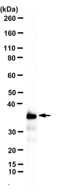
Western Blotting Analysis: A representative lot detected XPD in Western Blotting applications (Alekseev, S., et. al. (2017). Mol Cell. 65(3):504-514.e4).Immunocytochemistry Analysis: A representative lot detected XPD in Immunocytochmeistry applications (Vermeulen, W., et. al. (2000). Nat Genet. 26(3):307-13). .Immunoprecipitation Analysis: A representative lot immunoprecipitated XPD in Immunoprecipitation applications (Coin, F., et. al. (1998). Nat Genet. 20(2):184-8; Seroz, T., et. al. (2000). Nucleic Acids Res. 28(22):4506-13).
Classification
Monoclonal
Form
Purified
Regulatory Status
RUO
Target Species
Human
Gene Alias
General transcription and DNA repair factor IIH helicase subunit XPD;EC: 3.6.4.12;TFIIH subunit XPD;Basic transcription factor 2 80 kDa subunit;BTF2 p80;CXPD;DNA excision repair protein ERCC-2;DNA repair protein complementing XP-D cells;TFIIH basal transcription factor complex 80 kDa subunit;TFIIH 80 kDa subunit;TFIIH p80;Xeroderma pigmentosum group D-complementing protein
Gene Symbols
ERCC2;XPD;XPDC
Isotype
IgG1 κ
Purification Method
Protein G purified
Test Specificity
Clone 22TF2-2F6 detects General transcription and DNA repair factor IIH helicase subunit XPD (ERCC2) in human cells. It targets an epitope with in 32 amino acids from the C-terminal region.
Clone
22TF2-2F6
Applications
Immunocytochemistry, Immunoprecipitation, Western Blot
Conjugate
Unconjugated
Host Species
Mouse
Research Discipline
Epigenetics & Nuclear Function
Formulation
Purified mouse monoclonal antibody IgG1 in buffer containing 0.1 M Tris-Glycine (pH 7.4), 150 mM NaCl with 0.05% sodium azide.
Gene ID (Entrez)
NP_000391.1
Immunogen
Ovalbumin-conjugated linear peptide corresponding to 32 amino acids from the C-terminal region of human General transcription and DNA repair factor IIH helicase subunit XPD (XPD/ERCC2).
Primary or Secondary
Primary
Content And Storage
Stable for 1 year at 2-8°C from date of receipt.
Description
- Anti-XPD, clone 22TF2-2F6, Cat
- No
- MABE1819, is a highly specific mouse monoclonal antibody that targets human General transcription and DNA repair factor IIH helicase subunit XPD and has been tested for use in Immunocytochemistry, Immunoprecipitation, and Western Blotting
- General transcription and DNA repair factor IIH helicase subunit XPD (UniProt: P18074; also known as EC: 3.6.4.12, TFIIH subunit XPD, Basic transcription factor 2 80 kDa subunit, BTF2 p80, CXPD, DNA excision repair protein ERCC-2, DNA repair protein complementing XP-D cells, TFIIH basal transcription factor complex 80 kDa subunit, TFIIH 80 kDa subunit, TFIIH p80, Xeroderma pigmentosum group D-complementing protein) is encoded by the ERCC2 (also known as XPD, XPDC) gene (Gene ID: 2068) in human
- XPD is an ATP-dependent 5'-3' DNA helicase component of the general transcription and DNA repair factor IIH (TFIIH) core complex that is involved in general and transcription-coupled nucleotide excision repair (NER) of damaged DNA
- Its ATP-binding region is localized to amino acids 7-283 and its nuclear localization is in amino acids 682-695
- In NER, TFIIH acts by opening DNA around the lesion to allow the excision of the damaged oligonucleotide and its replacement by a new DNA fragment
- In transcription, TFIIH plays an essential role in transcription initiation
- When the pre-initiation complex has been established, TFIIH is required for promoter opening and promoter escape
- Phosphorylation of the C-terminal tail of the largest subunit of RNA polymerase II by CDK-activating kinase (CAK) complex controls the initiation of transcription
- XPD acts by forming a bridge between CAK and the core-TFIIH complex
- Mutations in ERCC2 gene are known to cause Xeroderma pigmentosum complementation group D that is characterized by solar hypersensitivity of the skin, high predisposition for developing cancers on areas exposed to sunlight and, in some cases, neurological abnormalities
- The skin develops marked freckling and other pigmentation abnormalities.
Compare Similar Items
Show Difference

Antigen: XPD
Dilution: Western Blotting Analysis: A representative lot detected XPD in Western Blotting applications (Alekseev, S., et. al. (2017). Mol Cell. 65(3):504-514.e4).Immunocytochemistry Analysis: A representative lot detected XPD in Immunocytochmeistry applications (Vermeulen, W., et. al. (2000). Nat Genet. 26(3):307-13). .Immunoprecipitation Analysis: A representative lot immunoprecipitated XPD in Immunoprecipitation applications (Coin, F., et. al. (1998). Nat Genet. 20(2):184-8; Seroz, T., et. al. (2000). Nucleic Acids Res. 28(22):4506-13).
Classification: Monoclonal
Form: Purified
Regulatory Status: RUO
Target Species: Human
Gene Alias: General transcription and DNA repair factor IIH helicase subunit XPD;EC: 3.6.4.12;TFIIH subunit XPD;Basic transcription factor 2 80 kDa subunit;BTF2 p80;CXPD;DNA excision repair protein ERCC-2;DNA repair protein complementing XP-D cells;TFIIH basal transcription factor complex 80 kDa subunit;TFIIH 80 kDa subunit;TFIIH p80;Xeroderma pigmentosum group D-complementing protein
Gene Symbols: ERCC2;XPD;XPDC
Isotype: IgG1 κ
Purification Method: Protein G purified
Test Specificity: Clone 22TF2-2F6 detects General transcription and DNA repair factor IIH helicase subunit XPD (ERCC2) in human cells. It targets an epitope with in 32 amino acids from the C-terminal region.
Clone: 22TF2-2F6
Applications: Immunocytochemistry, Immunoprecipitation, Western Blot
Conjugate: Unconjugated
Host Species: Mouse
Research Discipline: Epigenetics & Nuclear Function
Formulation: Purified mouse monoclonal antibody IgG1 in buffer containing 0.1 M Tris-Glycine (pH 7.4), 150 mM NaCl with 0.05% sodium azide.
Gene ID (Entrez): NP_000391.1
Immunogen: Ovalbumin-conjugated linear peptide corresponding to 32 amino acids from the C-terminal region of human General transcription and DNA repair factor IIH helicase subunit XPD (XPD/ERCC2).
Primary or Secondary: Primary
Content And Storage: Stable for 1 year at 2-8°C from date of receipt.
Antigen: AsCpf1
Dilution: Western Blotting Analysis: A 1:1,000 dilution from a representative lot detected AsCpf1 in HEK293 cells expressing HA-tagged AsCpf1 (Courtesy of Stefan Schuchner, Ph.D. and Egon Ogris, M.D., Medical University of Vienna, Austria).
Classification: Monoclonal
Form: Purified
Regulatory Status: RUO
Target Species: Bacteria
Gene Alias: EC 3.1.27.2;AsCpf1;CRISPR-associated endonuclease Cpf1
Gene Symbols: Cas12a;Cpf1
Isotype: IgG2a κ
Purification Method: Protein G purified
Test Specificity: Clone 3D3-F7 specifically detects CRISPR-associated endonuclease Cas12a.
Clone: 3D3-F7
Applications: Western Blot
Conjugate: Unconjugated
Host Species: Mouse
Research Discipline: Epigenetics & Nuclear Function
Formulation: Purified mouse monoclonal antibody IgG2a in buffer containing 0.1 M Tris-Glycine (pH 7.4), 150 mM NaCl with 0.05% sodium azide.
Gene ID (Entrez): __
Immunogen: His-tagged recombinant fragment corresponding to 197 amino acids from the C-terminal region of Acidaminococcus Cas12a.
Primary or Secondary: Primary
Content And Storage: Stable for 1 year at 2-8°C from date of receipt.
Antigen: beta-Catenin
Dilution: Western Blotting Analysis: A representative lot detected beta-Catenin in Western Blotting applications (Hoffmeyer, K., et. al. (2017). Cell Rep. 18(12):2815-2824).Immunoprecipitation Analysis: A representative lot immunoprecipitated beta-Catenin in Immunoprecipitation applications (Hoffmeyer, K., et. al. (2017). Cell Rep. 18(12):2815-2824).Dot Blot Analysis: A representative lot detected beta-Catenin in Dot Blot applications (Hoffmeyer, K., et. al. (2017). Cell Rep. 18(12):2815-2824). Chromatin Immunoprecipitation Analysis (ChIP): A representative lot immunoprecipitated beta-Catenin in Chromatin Immunoprecipitation applications (Hoffmeyer, K., et. al. (2017). Cell Rep. 18(12):2815-2824).
Classification: Monoclonal
Form: Purified
Regulatory Status: RUO
Target Species: Human
Gene Alias: beta-Catenin 1;Catenin beta-1
Gene Symbols: CTNNB1;CTNNB;OK/sw-cl.35;PRO2286
Isotype: IgG1 κ
Purification Method: Protein G purified
Test Specificity: This rat monoclonal antibody detects beta-catenin 1 unmodified and methylated or acetylated on lysine 49. It targets an epitope with in 19 amino acids from the N-terminal region.
Clone: mAb-Cat1
Applications: ChIP Assay, Dot Blot, Immunoprecipitation, Western Blot
Conjugate: Unconjugated
Host Species: Rat
Research Discipline: Epigenetics & Nuclear Function
Formulation: Purified rat monoclonal antibody IgG1 in buffer containing 0.1 M Tris-Glycine (pH 7.4), 150 mM NaCl with 0.05% sodium azide.
Gene ID (Entrez): NP_001091679.1
Immunogen: BSA-conjugated linear peptide corresponding to 19 amino acids from the N-terminal region of human beta-catenin 1.
Primary or Secondary: Primary
Content And Storage: Stable for 1 year at 2-8°C from date of receipt.