Desmoplakin, Mouse anti-Human, Clone: 20B6, Millipore Sigma™
Mouse Monoclonal Antibody
Manufacturer: Fischer Scientific
The price for this product is unavailable. Please request a quote
Antigen
Desmoplakin
Dilution
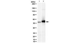
Western Blotting Analysis: A representative lot detected Desmoplakin in Western Blotting applications (Sobolik-Delmaire, T., et. al. (2006). J Biol Chem. 281(25):16962-70; Hall, C., et. al. (2009). Cell Commun Adhes. 16(1-3):15-27; Roberts, B.J., et. al. (2011). Exp Cell Res. 317(20):2814-22).Immunocytochemistry Analysis: A representative lot detected Desmoplakin in Immunocytochemistry applications (Roberts, B.J., et. al. (2011). Exp Cell Res. 317(20):2814-22; Sobolik-Delmaire, T., et. al. (2006). J Biol Chem. 281(25):16962-70; Roberts, B.J., et. al. (2016). J Biol Chem. 291(48):24857-24865).
Classification
Monoclonal
Form
Purified
Regulatory Status
RUO
Target Species
Human
Gene Alias
DP;250/210 kDa paraneoplastic pemphigus antigen
Gene Symbols
DSP
Isotype
IgG1 κ
Purification Method
Protein G purified
Test Specificity
Clone 20B6 detects human Desmoplankin. It targets an epitope with in the C-terminal half.
Clone
20B6
Applications
Immunocytochemistry, Western Blot
Conjugate
Unconjugated
Host Species
Mouse
Research Discipline
Cell Structure
Formulation
Purified mouse monoclonal antibody IgG1 in buffer containing 0.1 M Tris-Glycine (pH 7.4), 150 mM NaCl with 0.05% sodium azide.
Gene ID (Entrez)
NP_001008844
Immunogen
His-tagged recombinant fragment corresponding to 863 amino acids from the C-terminal half of human Desmoplakin.
Primary or Secondary
Primary
Content And Storage
Stable for 1 year at 2-8°C from date of receipt.
Description
- Anti-Desmoplakin, clone 20B6, Cat
- No
- MABT1492, is a mouse monoclonal antibody that detects human Desmoplakin and has been tested for use in Immunocytochemistry and Western Blotting
- Desmoplakin (UniProt: P15924; also known as DP, 250/210 kDa paraneoplastic pemphigus antigen) is encoded by the DSP gene (Gene ID:1832) in human
- Desmoplakin is a major high molecular weight homodimeric protein that is involved in the organization of the desmosomal cadherin-plakoglobin complexes into discrete plasma membrane domains and in the anchoring of intermediate filaments to the desmosomes, which are prominent adhesive junctions present between many epithelial cells and cardiomyocytes
- Desmoplakin is the most abundant of the desmosomal proteins and plays a key role in tissue morphogenesis and maintaining the integrity of tissue
- Desmoplakin is composed of an N-terminal plakin domain, a central alpha-helical coiled-coil rod domain, and a C-terminal intermediate filament-binding domain
- The N-terminal domain is required for desmosome association and the C-terminal binds to desmin
- Three isoforms for Desmoplakin have been reported that are produced by alternative splicing
- Desmoplakin 2 and 3 lack most of the central rod domain
- Desmoplakin 1 is the dominant isoform in the cardiac tissue
- Desmoplakin is phosphorylated by protein kinase A on serine 2849 and this phosphorylation affects its association with epidermal, simple cytokeratins, and VIM intermediate filaments
- Mutations in DSP gene have been linked to DCWHK (Cardiomyopathy, dilated, with woolly hair and keratoderma) that is characterized by a generalized striate keratoderma particularly affecting the palmoplantar epidermis, woolly hair, and dilated left ventricular cardiomyopathy
- Mice deficient in Desmoplakin perish around the time of implantation (E6.5) and show fewer desmosomes.
Compare Similar Items
Show Difference

Antigen: Desmoplakin
Dilution: Western Blotting Analysis: A representative lot detected Desmoplakin in Western Blotting applications (Sobolik-Delmaire, T., et. al. (2006). J Biol Chem. 281(25):16962-70; Hall, C., et. al. (2009). Cell Commun Adhes. 16(1-3):15-27; Roberts, B.J., et. al. (2011). Exp Cell Res. 317(20):2814-22).Immunocytochemistry Analysis: A representative lot detected Desmoplakin in Immunocytochemistry applications (Roberts, B.J., et. al. (2011). Exp Cell Res. 317(20):2814-22; Sobolik-Delmaire, T., et. al. (2006). J Biol Chem. 281(25):16962-70; Roberts, B.J., et. al. (2016). J Biol Chem. 291(48):24857-24865).
Classification: Monoclonal
Form: Purified
Regulatory Status: RUO
Target Species: Human
Gene Alias: DP;250/210 kDa paraneoplastic pemphigus antigen
Gene Symbols: DSP
Isotype: IgG1 κ
Purification Method: Protein G purified
Test Specificity: Clone 20B6 detects human Desmoplankin. It targets an epitope with in the C-terminal half.
Clone: 20B6
Applications: Immunocytochemistry, Western Blot
Conjugate: Unconjugated
Host Species: Mouse
Research Discipline: Cell Structure
Formulation: Purified mouse monoclonal antibody IgG1 in buffer containing 0.1 M Tris-Glycine (pH 7.4), 150 mM NaCl with 0.05% sodium azide.
Gene ID (Entrez): NP_001008844
Immunogen: His-tagged recombinant fragment corresponding to 863 amino acids from the C-terminal half of human Desmoplakin.
Primary or Secondary: Primary
Content And Storage: Stable for 1 year at 2-8°C from date of receipt.

Antigen: PD2 (PAF1)
Dilution: Immunocytochemistry Analysis: A representative lot detected PD2 (PAF1) in Immunocytochemistry applications (Dey, P., et. al. (2014). Oncotarget. 5(12):4480-91; Moniaux, N., et. al. (2006). Oncogene. 25(23):3247-57).Immunoprecipitation Analysis: A representative lot immunoprecipitated PD2 (PAF1) in Immunoprecipitation applications (Moniaux, N., et. al. (2006). Oncogene. 25(23):3247-57).ELISA Analysis: A representative lot detected PD2 (PAF1) in ELISA applications (Moniaux, N., et. al. (2006). Oncogene. 25(23):3247-57).Immunohistochemistry Analysis: A representative lot detected PD2 (PAF1) in Immunohistochemistry applications (Dey, P., et. al. (2014). Oncotarget. 5(12):4480-91).Western Blotting Analysis: A representative lot detected PD2 (PAF1) in Western Blotting applications (Moniaux, N., et. al. (2006). Oncogene. 25(23):3247-57).
Classification: Monoclonal
Form: Purified
Regulatory Status: RUO
Target Species: Human
Gene Alias: Pancreatic differentiation protein 2;RNA polymerase II-associated factor 1 homolog;hPAF1
Gene Symbols: PAF1;PD2
Isotype: IgG1 κ
Purification Method: Protein G purified
Test Specificity: Clone F169-3B2 detects Pancreatic differentiation protein 2 (PD2) in human. It targets an epitope with in 22 amino acids from the C-terminal half.
Clone: F169-3B2
Applications: ELISA, Immunocytochemistry, Immunohistochemistry, Immunoprecipitation, Western Blot
Conjugate: Unconjugated
Host Species: Mouse
Research Discipline: Epigenetics & Nuclear Function
Formulation: Purified mouse monoclonal antibody IgG1 in PBS without azide.
Gene ID (Entrez): NP_061961
Immunogen: KLH-conjugated linear peptide corresponding to 22 amino acids from the C-terminal half of human Pancreatic differentiation protein 2 (PD2).
Primary or Secondary: Primary
Content And Storage: Stable for 1 year at -20°C from date of receipt. Handling Recommendations: Upon receipt and prior to removing the cap, centrifuge the vial and gently mix the solution. Aliquot into microcentrifuge tubes and store at -20°C. Avoid repeated freeze/thaw cycles, which may damage IgG and affect product performance.

Antigen: P40 (TP63)
Dilution: __
Classification: Monoclonal
Form: Purified
Regulatory Status: RUO
Target Species: Human
Gene Alias: Tumor protein 63;p63;Chronic ulcerative stomatitis protein;CUSP;Keratinocyte transcription factor KET;Transformation-related protein 63;TP63;Tumor protein p73-like;p73L;P51
Gene Symbols: P63;KET;P63;P73H;P73L;TP73L
Isotype: IgG1 κ
Purification Method: Protein G purified
Test Specificity: Clone 11H1 detects human Tumor protein 63 (P40). It targets an epitope with in the first 205 amino acids from the N-terminal region.
Clone: 11H1
Applications: Western Blot
Conjugate: Unconjugated
Host Species: Mouse
Research Discipline: Epigenetics & Nuclear Function
Formulation: Purified mouse monoclonal antibody IgG1 in buffer containing 0.1 M Tris-Glycine (pH 7.4), 150 mM NaCl with 0.05% sodium azide.
Gene ID (Entrez): NP_001108452
Immunogen: MBP-conjugated recombinant fragment corresponding to the first 205 amino acids from human Tumor protein 63.
Primary or Secondary: Primary
Content And Storage: Stable for 1 year at 2-8°C from date of receipt.