Cytokeratin 10 Antibody (DE-K10) - Azide and BSA Free, Novus Biologicals™
Manufacturer: Fischer Scientific
Select a Size
| Pack Size | SKU | Availability | Price |
|---|---|---|---|
| Each of 1 | NB254402100-Each-of-1 | In Stock | ₹ 57,494.00 |
NB254402100 - Each of 1
In Stock
Quantity
1
Base Price: ₹ 57,494.00
GST (18%): ₹ 10,348.92
Total Price: ₹ 67,842.92
Antigen
Cytokeratin 10
Classification
Monoclonal
Concentration
1 mg/mL
Dilution
Flow Cytometry 0.5 - 1 ug/million cells, Immunocytochemistry/Immunofluorescence 0.5 - 1 ug/ml, Immunohistochemistry-Paraffin 0.1 - 0.2 ug/ml, CyTOF-ready
Gene Alias
BCIE, BIE, CK10, CK-10, cytokeratin 10, Cytokeratin-10, EHK, K10keratosis palmaris et plantaris, keratin 10, Keratin 10 (epidermolytic hyperkeratosis; keratosis palmaris et plantaris), keratin, type I cytoskeletal 10, keratin-10, KPP
Host Species
Mouse
Molecular Weight of Antigen
56.5 kDa
Quantity
100 μg
Research Discipline
Cytoskeleton Markers
Gene ID (Entrez)
3858
Target Species
Human, Canine, Feline
Form
Purified
Applications
Flow Cytometry, Immunocytochemistry, Immunofluorescence, Immunohistochemistry (Paraffin), CyTOF
Clone
DE-K10
Conjugate
Unconjugated
Formulation
PBS with No Preservative
Gene Symbols
KRT10
Immunogen
Cytoskeletal preparation extracted from human ectocervical epithelium
Purification Method
Protein A or G purified
Regulatory Status
RUO
Primary or Secondary
Primary
Test Specificity
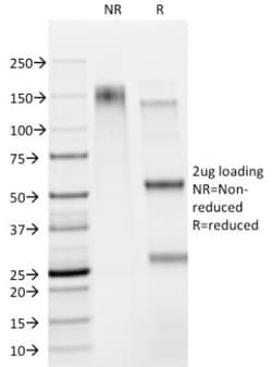
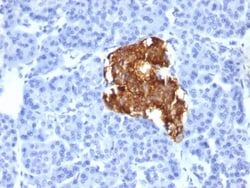
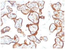
This MAb recognizes a protein of 56.5kDa, identified as cytokeratin 10 (CK10). CK10 is expressed in all suprabasal layers of the epidermis. In the epidermis, expression of CK10 strictly parallels the extent of differentiation; it is absent in the basal layer, appears in the first suprabasal layers and increases in concentration towards the granular layer. However, CK10 is rarely detected in early stages of vulvar squamous carcinomas (tumors less than 2 cm, clinical stage I) regardless of the tumor grade. In larger and more advanced tumors (greater than 2 cm, clinical stages II and III), CK10 is detected very frequently. Expression of CK10 is related to maturation of malignant keratinocytes, being preferentially detected in more-differentiated parts.
Content And Storage
Store at -20 to -80C. Avoid freeze-thaw cycles.
Isotype
IgG1 κ
Related Products


Description
- Cytokeratin 10 Monoclonal specifically detects Cytokeratin 10 in Human, Canine, Feline samples
- It is validated for Immunohistochemistry, Immunohistochemistry-Paraffin.
Compare Similar Items
Show Difference
Antigen: Cytokeratin 10
Classification: Monoclonal
Concentration: 1 mg/mL
Dilution: Flow Cytometry 0.5 - 1 ug/million cells, Immunocytochemistry/Immunofluorescence 0.5 - 1 ug/ml, Immunohistochemistry-Paraffin 0.1 - 0.2 ug/ml, CyTOF-ready
Gene Alias: BCIE, BIE, CK10, CK-10, cytokeratin 10, Cytokeratin-10, EHK, K10keratosis palmaris et plantaris, keratin 10, Keratin 10 (epidermolytic hyperkeratosis; keratosis palmaris et plantaris), keratin, type I cytoskeletal 10, keratin-10, KPP
Host Species: Mouse
Molecular Weight of Antigen: 56.5 kDa
Quantity: 100 μg
Research Discipline: Cytoskeleton Markers
Gene ID (Entrez): 3858
Target Species: Human, Canine, Feline
Form: Purified
Applications: Flow Cytometry, Immunocytochemistry, Immunofluorescence, Immunohistochemistry (Paraffin), CyTOF
Clone: DE-K10
Conjugate: Unconjugated
Formulation: PBS with No Preservative
Gene Symbols: KRT10
Immunogen: Cytoskeletal preparation extracted from human ectocervical epithelium
Purification Method: Protein A or G purified
Regulatory Status: RUO
Primary or Secondary: Primary
Test Specificity: This MAb recognizes a protein of 56.5kDa, identified as cytokeratin 10 (CK10). CK10 is expressed in all suprabasal layers of the epidermis. In the epidermis, expression of CK10 strictly parallels the extent of differentiation; it is absent in the basal layer, appears in the first suprabasal layers and increases in concentration towards the granular layer. However, CK10 is rarely detected in early stages of vulvar squamous carcinomas (tumors less than 2 cm, clinical stage I) regardless of the tumor grade. In larger and more advanced tumors (greater than 2 cm, clinical stages II and III), CK10 is detected very frequently. Expression of CK10 is related to maturation of malignant keratinocytes, being preferentially detected in more-differentiated parts.
Content And Storage: Store at -20 to -80C. Avoid freeze-thaw cycles.
Isotype: IgG1 κ
Antigen: Cytokeratin 10
Classification: Monoclonal
Concentration: 1 mg/mL
Dilution: Flow Cytometry 0.5 - 1 ug/million cells, Immunocytochemistry/Immunofluorescence 0.5 - 1 ug/ml, Immunohistochemistry-Paraffin 0.1 - 0.2 ug/ml, CyTOF-ready
Gene Alias: BCIE, BIE, CK10, CK-10, cytokeratin 10, Cytokeratin-10, EHK, K10keratosis palmaris et plantaris, keratin 10, Keratin 10 (epidermolytic hyperkeratosis; keratosis palmaris et plantaris), keratin, type I cytoskeletal 10, keratin-10, KPP
Host Species: Mouse
Molecular Weight of Antigen: 56.5 kDa
Quantity: 200 μg
Research Discipline: Cytoskeleton Markers
Gene ID (Entrez): 3858
Target Species: Human, Canine, Feline
Form: Purified
Applications: Flow Cytometry, Immunocytochemistry, Immunofluorescence, Immunohistochemistry (Paraffin), CyTOF
Clone: DE-K10
Conjugate: Unconjugated
Formulation: PBS with No Preservative
Gene Symbols: KRT10
Immunogen: Cytoskeletal preparation extracted from human ectocervical epithelium
Purification Method: Protein A or G purified
Regulatory Status: RUO
Primary or Secondary: Primary
Test Specificity: This MAb recognizes a protein of 56.5kDa, identified as cytokeratin 10 (CK10). CK10 is expressed in all suprabasal layers of the epidermis. In the epidermis, expression of CK10 strictly parallels the extent of differentiation; it is absent in the basal layer, appears in the first suprabasal layers and increases in concentration towards the granular layer. However, CK10 is rarely detected in early stages of vulvar squamous carcinomas (tumors less than 2 cm, clinical stage I) regardless of the tumor grade. In larger and more advanced tumors (greater than 2 cm, clinical stages II and III), CK10 is detected very frequently. Expression of CK10 is related to maturation of malignant keratinocytes, being preferentially detected in more-differentiated parts.
Content And Storage: Store at -20 to -80C. Avoid freeze-thaw cycles.
Isotype: IgG1 κ
Antigen: PD-ECGF/Thymidine Phosphorylase
Classification: Monoclonal
Concentration: 1 mg/mL
Dilution: Western Blot 0.5 - 1 ug/ml, Immunoprecipitation 0.5 - 1 ug, Immunohistochemistry-Paraffin 1 - 2 ug/ml
Gene Alias: EC 2.4.2, EC 2.4.2.4, ECGF, ECGF1TP, endothelial cell growth factor 1 (platelet-derived), Gliostatin, hPD-ECGF, MEDPS1, MNGIE, MTDPS1, PDECGF, PD-ECGF, Platelet-derived endothelial cell growth factor, TdRPase, thymidine phosphorylase
Host Species: Mouse
Molecular Weight of Antigen: 55 kDa
Quantity: 100 μg
Research Discipline: Angiogenesis
Gene ID (Entrez): 1890
Target Species: Human, Mouse, Rat
Form: Purified
Applications: Western Blot, Immunoprecipitation, Immunohistochemistry (Paraffin)
Clone: SPM322
Conjugate: Unconjugated
Formulation: PBS with No Preservative
Gene Symbols: TYMP
Immunogen: Recombinant full-length human Thymidine Phosphorylase (TP/ PD-ECGF) protein
Purification Method: Protein A or G purified
Regulatory Status: RUO
Primary or Secondary: Primary
Test Specificity: __
Content And Storage: Store at -20 to -80C. Avoid freeze-thaw cycles.
Isotype: IgG1