PMEL17/SILV Antibody (NKI-beteb), Novus Biologicals™
Manufacturer: Novus Biologicals
Select a Size
| Pack Size | SKU | Availability | Price |
|---|---|---|---|
| Each of 1 | NBP229407-Each-of-1 | In Stock | ₹ 46,636.00 |
NBP229407 - Each of 1
In Stock
Quantity
1
Base Price: ₹ 46,636.00
GST (18%): ₹ 8,394.48
Total Price: ₹ 55,030.48
Antigen
PMEL17/SILV
Classification
Monoclonal
Concentration
0.2 mg/ml
Dilution
Immunohistochemistry, Immunohistochemistry-Paraffin 1-2 ug/ml, Protein Array, Flow (Intracellular)
Gene Alias
D12S53EP1, gp100, ME20, ME20-M, melanocyte protein mel 17, Melanocyte protein Pmel 17, Melanocytes lineage-specific antigen GP100, Melanoma-associated ME20 antigen, melanosomal matrix protein17, PMEL17P100, premelanosome proteinME20M, SI, SIL, silver (mouse homolog) like, silver homolog (mouse), Silver locus protein homolog, silver, mouse, homolog of, SILVPmel17
Host Species
Mouse
Purification Method
Protein A or G purified
Regulatory Status
RUO
Gene ID (Entrez)
6490
Target Species
Human, Equine
Form
Purified
Applications
Immunohistochemistry, Immunohistochemistry (Paraffin), Peptide Array, Flow Cytometry, Immunohistochemistry (Frozen)
Clone
NKI-beteb
Conjugate
Unconjugated
Gene Accession No.
P40967
Gene Symbols
PMEL
Immunogen
Lymph node pigmented melanoma metastases extract was used as the immunogen for the gp100 NKI-beteb antibody
Quantity
0.1 mg
Primary or Secondary
Primary
Test Specificity
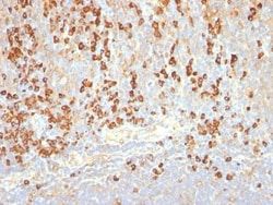
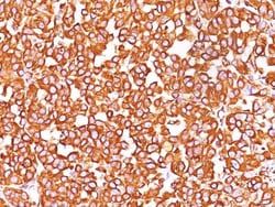
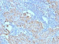
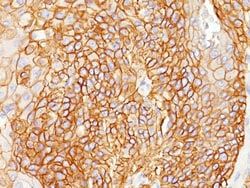
By immunohistochemistry, it specifically recognizes a protein in melanocytes and melanomas. This monoclonal antibody reacts with junctional and blue nevus cells and variably with fetal and neonatal melanocytes. Intradermal nevi, normal adult melanocytes, and non-melanocytic cells are negative. It does not stain tumor cells of epithelial, lymphoid, glial, or mesenchymal origin. This Mab labels formalin-fixed, paraffin-embedded melanomas and other tumors showing melanocytic differentiation.
Content And Storage
Store at 4C.
Isotype
IgG2b κ
Related Products


Description
- PMEL17/SILV Monoclonal specifically detects PMEL17/SILV in Human, Equine samples
- It is validated for Flow Cytometry, Immunohistochemistry, Immunohistochemistry-Paraffin, Protein Array, Flow (Intracellular).
Compare Similar Items
Show Difference

Antigen: PMEL17/SILV
Classification: Monoclonal
Concentration: 0.2 mg/ml
Dilution: Immunohistochemistry, Immunohistochemistry-Paraffin 1-2 ug/ml, Protein Array, Flow (Intracellular)
Gene Alias: D12S53EP1, gp100, ME20, ME20-M, melanocyte protein mel 17, Melanocyte protein Pmel 17, Melanocytes lineage-specific antigen GP100, Melanoma-associated ME20 antigen, melanosomal matrix protein17, PMEL17P100, premelanosome proteinME20M, SI, SIL, silver (mouse homolog) like, silver homolog (mouse), Silver locus protein homolog, silver, mouse, homolog of, SILVPmel17
Host Species: Mouse
Purification Method: Protein A or G purified
Regulatory Status: RUO
Gene ID (Entrez): 6490
Target Species: Human, Equine
Form: Purified
Applications: Immunohistochemistry, Immunohistochemistry (Paraffin), Peptide Array, Flow Cytometry, Immunohistochemistry (Frozen)
Clone: NKI-beteb
Conjugate: Unconjugated
Gene Accession No.: P40967
Gene Symbols: PMEL
Immunogen: Lymph node pigmented melanoma metastases extract was used as the immunogen for the gp100 NKI-beteb antibody
Quantity: 0.1 mg
Primary or Secondary: Primary
Test Specificity: By immunohistochemistry, it specifically recognizes a protein in melanocytes and melanomas. This monoclonal antibody reacts with junctional and blue nevus cells and variably with fetal and neonatal melanocytes. Intradermal nevi, normal adult melanocytes, and non-melanocytic cells are negative. It does not stain tumor cells of epithelial, lymphoid, glial, or mesenchymal origin. This Mab labels formalin-fixed, paraffin-embedded melanomas and other tumors showing melanocytic differentiation.
Content And Storage: Store at 4C.
Isotype: IgG2b κ
Antigen: Cytokeratin 18
Classification: Monoclonal
Concentration: 0.2 mg/ml
Dilution: Western Blot 1-3 μg/mL, Simple Western 10 μg/mL, Flow Cytometry 0.5-1 μg/million cells, ELISA 1-5 μg/mL for coating, Immunohistochemistry, Immunocytochemistry/Immunofluorescence 1-2 μg/mL, Immunoprecipitation 1-2 μg/500 μg protein lysate, Immunohistochemistry-Paraffin 0.5-1.0 μg/mL
Gene Alias: Cell proliferation-inducing gene 46 protein, cell proliferation-inducing protein 46, CK-18, CYK18, cytokeratin 18, cytokeratin-18, K18, keratin 18, keratin, type I cytoskeletal 18, keratin-18
Host Species: Mouse
Purification Method: Protein A or G purified
Regulatory Status: RUO
Gene ID (Entrez): 3875
Target Species: Human, Bovine (Negative), Canine (Negative), Hamster (Negative), Mouse (Negative), Porcine (Negative), Rat (Negative)
Form: Purified
Applications: Western Blot, Flow Cytometry, ELISA, Immunohistochemistry, Immunocytochemistry, Immunofluorescence, Immunoprecipitation, Immunohistochemistry (Paraffin)
Clone: DA7
Conjugate: Unconjugated
Gene Accession No.: P05783
Gene Symbols: KRT18
Immunogen: Human breast cancer PMC 42 cells were used as immunogen to generate the Cytokeratin 18 (CK18) antibody.
Quantity: 0.1 mg
Primary or Secondary: Primary
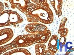
Test Specificity: This monoclonal antibody reacts with a wide variety of simple epithelia. It does not react with stratified squamous epithelia. It reacts with epithelial tumors of the gastrointestinal tract, lung, breast, pancreas, ovary, and thyroid. Cytokeratin 18, which belongs to the type A (acidic) subfamily of low molecular weight keratins, exists in combination with cytokeratin 8. It was reported that tissues from gastrointestinal tract are positive for both cytokeratin 8 and 18 but do not contain cytokeratin 14. Tissues from gastrointestinal tract, respiratory tract and urogenital tract, as well as endocrine and exocrine tissues and mesothelial cells are positive for cytokeratin 18.
Content And Storage: Store at 4C.
Isotype: IgG1 κ
Antigen: Cytokeratin, LMW
Classification: Monoclonal
Concentration: 0.2 mg/ml
Dilution: Western Blot 0.5-1 μg/mL, Flow Cytometry 0.5-1 μg/million cells, ELISA 1-5 μg/mL for coating, Immunohistochemistry, Immunocytochemistry/Immunofluorescence 1-2 μg/mL, Immunohistochemistry-Paraffin 0.5-1 μg/mL, Immunohistochemistry-Frozen 0.5-1 μg/mL
Gene Alias: Cytokeratin LMW, cytokeratin-1B, keratin 77, keratin, type II cytoskeletal 1b, KRT77, type-II keratin Kb39
Host Species: Mouse
Purification Method: Protein A or G purified
Regulatory Status: RUO
Gene ID (Entrez): 374454
Target Species: Human, Mouse, Rat, Bovine, Canine, Chicken, Other, Primate, Rabbit
Form: Purified
Applications: Western Blot, Flow Cytometry, ELISA, Immunohistochemistry, Immunocytochemistry, Immunofluorescence, Immunohistochemistry (Paraffin), Immunohistochemistry (Frozen)
Clone: AE-1
Conjugate: Unconjugated
Gene Accession No.: P04264
Gene Symbols: KRT77
Immunogen: Human epidermal keratin (Uniprot: Q7Z794)
Quantity: 0.1 mg
Primary or Secondary: Primary
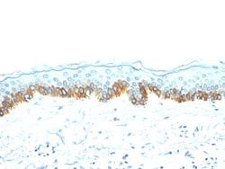
Test Specificity: This monoclonal antibody recognizes the 56.5kDa (CK10); 50kDa (CK14); 50kDa (CK15); 48kDa (CK16); 40kDa (CK19) keratins of the acidic (Type I or LMW) subfamily. Twenty human keratins are resolved with two-dimensional gel electrophoresis into acidic (pI, 48, 46, 45, and 40kDa. monoclonal antibody AE3 recognizes the 65-67, 64, 59, 58, 56, and 52kDa keratins of basic subfamily. Many studies have shown the usefulness of keratins as markers in cancer research and tumor diagnosis. AE1/AE3 is a broad spectrum anti pan-keratin antibody cocktail, which differentiates epithelial tumors from non-epithelial tumors e.g. squamous vs. adenocarcinoma of the lung, liver carcinoma, breast cancer, and esophageal cancer.
Content And Storage: Store at 4C.
Isotype: IgG1 κ