Endorepellin/Perlecan/Heparan Sulfate Proteoglycan Antibody (SPM255), Novus Biologicals™
Manufacturer: Fischer Scientific
The price for this product is unavailable. Please request a quote
Antigen
Endorepellin/Perlecan/Heparan Sulfate Proteoglycan
Classification
Monoclonal
Concentration
0.2mg/mL
Dilution
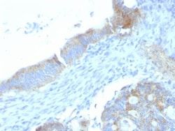
Flow Cytometry 0.5 - 1 ug/million cells in 0.1 ml, Immunohistochemistry-Paraffin 1 - 2 ug/ml, Immunofluorescence 0.5 - 1.0 ug/ml
Gene Alias
basement membrane-specific heparan sulfate proteoglycan core protein, endorepellin (domain V region), heparan sulfate proteoglycan 2, HSPG, perlecan, perlecan proteoglycan, PLCSchwartz-Jampel syndrome 1 (chondrodystrophic myotonia), PRCAN, SJA, SJS, SJS1
Host Species
Rat
Purification Method
Protein A or G purified
Regulatory Status
RUO
Gene ID (Entrez)
3339
Target Species
Human, Mouse, Porcine, Fish, Monkey, Primate
Form
Purified
Applications
Flow Cytometry, Immunohistochemistry (Paraffin), Immunofluorescence
Clone
SPM255
Conjugate
Unconjugated
Formulation
10mM PBS and 0.05% BSA with 0.05% Sodium Azide
Gene Symbols
HSPG2
Immunogen
Murine EHS laminin preparation
Quantity
0.2 mg
Primary or Secondary
Primary
Test Specificity
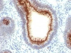
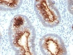
This MAb specifically precipitates heterogeneous material of high MW, identified as perlecan, a major heparan-sulfate proteoglycan (HSPG) within all basement membranes and cell surfaces. It does not cross-react with laminin, fibronectin, or dermatran sulfate proteoglycan. Because of perlecan s strategic location and ability to store and protect growth factors, it has been strongly implicated in the control of tumor cell growth and metastatic behavior. Perlecan possesses angiogenic and growth-promoting attributes primarily by acting as a co-receptor for basic fibroblast growth factor (FGF-2). Suppression of perlecan causes substantial inhibition of neoplastic growth and neovascularization. Thus, perlecan is a potent inducer of neoplasm growth and angiogenesis in vivo and therapeutic interventions targeting this key modulator of tumor progression may improve neoplastic treatment
Content And Storage
Store at 4C.
Isotype
IgG2a κ
Related Products


Description
- Ensure accurate, reproducible results in Flow Cytometry, Immunohistochemistry (Paraffin), Immunofluorescence Endorepellin/Perlecan/Heparan Sulfate Proteoglycan Monoclonal specifically detects Endorepellin/Perlecan/Heparan Sulfate Proteoglycan in Human, Mouse, Porcine, Bovine, Fish, Monkey samples
- It is validated for Flow Cytometry, Immunohistochemistry, Immunocytochemistry/Immunofluorescence, Immunohistochemistry-Paraffin, Immunofluorescence.
Compare Similar Items
Show Difference
Antigen: Endorepellin/Perlecan/Heparan Sulfate Proteoglycan
Classification: Monoclonal
Concentration: 0.2mg/mL
Dilution: Flow Cytometry 0.5 - 1 ug/million cells in 0.1 ml, Immunohistochemistry-Paraffin 1 - 2 ug/ml, Immunofluorescence 0.5 - 1.0 ug/ml
Gene Alias: basement membrane-specific heparan sulfate proteoglycan core protein, endorepellin (domain V region), heparan sulfate proteoglycan 2, HSPG, perlecan, perlecan proteoglycan, PLCSchwartz-Jampel syndrome 1 (chondrodystrophic myotonia), PRCAN, SJA, SJS, SJS1
Host Species: Rat
Purification Method: Protein A or G purified
Regulatory Status: RUO
Gene ID (Entrez): 3339
Target Species: Human, Mouse, Porcine, Fish, Monkey, Primate
Form: Purified
Applications: Flow Cytometry, Immunohistochemistry (Paraffin), Immunofluorescence
Clone: SPM255
Conjugate: Unconjugated
Formulation: 10mM PBS and 0.05% BSA with 0.05% Sodium Azide
Gene Symbols: HSPG2
Immunogen: Murine EHS laminin preparation
Quantity: 0.2 mg
Primary or Secondary: Primary
Test Specificity: This MAb specifically precipitates heterogeneous material of high MW, identified as perlecan, a major heparan-sulfate proteoglycan (HSPG) within all basement membranes and cell surfaces. It does not cross-react with laminin, fibronectin, or dermatran sulfate proteoglycan. Because of perlecan s strategic location and ability to store and protect growth factors, it has been strongly implicated in the control of tumor cell growth and metastatic behavior. Perlecan possesses angiogenic and growth-promoting attributes primarily by acting as a co-receptor for basic fibroblast growth factor (FGF-2). Suppression of perlecan causes substantial inhibition of neoplastic growth and neovascularization. Thus, perlecan is a potent inducer of neoplasm growth and angiogenesis in vivo and therapeutic interventions targeting this key modulator of tumor progression may improve neoplastic treatment
Content And Storage: Store at 4C.
Isotype: IgG2a κ

Antigen: Mucin 5AC
Classification: Monoclonal
Concentration: 0.2 mg/mL
Dilution: Flow Cytometry 0.5 - 1 ug/million cells in 0.1 ml, Immunohistochemistry-Frozen 0.5 - 1.0 ug/ml, Immunofluorescence 1 - 2 ug/ml
Gene Alias: gastric mucin, leB, lewis B blood group antigen, major airway glycoprotein, MUC5, mucin 5, subtypes A and C, tracheobronchial/gastric, mucin 5AC, oligomeric mucus/gel-forming, mucin 5AC, oligomeric mucus/gel-forming pseudogene, mucin-5 subtype AC, tracheobronchial, mucin-5AC, TBM, tracheobronchial mucin
Host Species: Mouse
Purification Method: Protein A or G purified
Regulatory Status: RUO
Gene ID (Entrez): 4586
Target Species: Human, Mouse, Rat, Chicken, Feline, Monkey, Rabbit, Porcine (Negative)
Form: Purified
Applications: Flow Cytometry, Immunohistochemistry (Frozen), Immunofluorescence
Clone: 1-13M1
Conjugate: Unconjugated
Formulation: 10mM PBS and 0.05% BSA with 0.05% Sodium Azide
Gene Symbols: MUC5AC
Immunogen: M1 mucin preparation from the fluid of an ovarian mucinous cyst belonging to an O Le(a-b) patient.
Quantity: 0.02 mg
Primary or Secondary: Primary
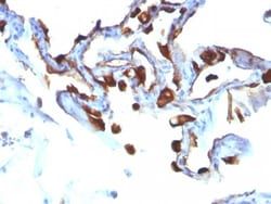
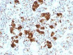
Test Specificity: This MAb recognizes the peptide core of gastric mucin M1 (recently identified as Mucin 5AC).Its epitope is located in the peptide core of MUC5AC. Its epitope is destroyed by beta-mercaptoethanol but not by periodate treatment. MAb 1-13M1 pairs with MAb 9-13M1 to measure MUC5AC protein by ELISA. This mucin is present in primary ovarian mucinous cancer but usually absent in colorectal adenocarcinoma, thus showing an expression pattern opposite to MUC2. Together with a panel of antibodies, Anti-MUC5AC may be useful for differential identification of primary mucinous ovarian tumors from colon adenocarcinoma metastatic to the ovary. MUC5AC antibodies may also be useful for identification of intestinal metaplasia as well as in the identification of pancreatic carcinoma and pre-cancerous changes vs. normal pancreas.
Content And Storage: Store at 4C.
Isotype: IgG1 κ
Antigen: Mucin 5AC
Classification: Monoclonal
Concentration: 0.2 mg/mL
Dilution: Flow Cytometry 0.5 - 1 ug/million cells in 0.1 ml, Immunohistochemistry-Frozen 0.5 - 1.0 ug/ml, Immunofluorescence 1 - 2 ug/ml
Gene Alias: gastric mucin, leB, lewis B blood group antigen, major airway glycoprotein, MUC5, mucin 5, subtypes A and C, tracheobronchial/gastric, mucin 5AC, oligomeric mucus/gel-forming, mucin 5AC, oligomeric mucus/gel-forming pseudogene, mucin-5 subtype AC, tracheobronchial, mucin-5AC, TBM, tracheobronchial mucin
Host Species: Mouse
Purification Method: Protein A or G purified
Regulatory Status: RUO
Gene ID (Entrez): 4586
Target Species: Human, Mouse, Rat, Chicken, Feline, Monkey, Rabbit, Porcine (Negative)
Form: Purified
Applications: Flow Cytometry, Immunohistochemistry (Frozen), Immunofluorescence
Clone: 1-13M1
Conjugate: Unconjugated
Formulation: 10mM PBS and 0.05% BSA with 0.05% Sodium Azide
Gene Symbols: MUC5AC
Immunogen: M1 mucin preparation from the fluid of an ovarian mucinous cyst belonging to an O Le(a-b) patient.
Quantity: 0.1 mg
Primary or Secondary: Primary
Test Specificity: This MAb recognizes the peptide core of gastric mucin M1 (recently identified as Mucin 5AC).Its epitope is located in the peptide core of MUC5AC. Its epitope is destroyed by beta-mercaptoethanol but not by periodate treatment. MAb 1-13M1 pairs with MAb 9-13M1 to measure MUC5AC protein by ELISA. This mucin is present in primary ovarian mucinous cancer but usually absent in colorectal adenocarcinoma, thus showing an expression pattern opposite to MUC2. Together with a panel of antibodies, Anti-MUC5AC may be useful for differential identification of primary mucinous ovarian tumors from colon adenocarcinoma metastatic to the ovary. MUC5AC antibodies may also be useful for identification of intestinal metaplasia as well as in the identification of pancreatic carcinoma and pre-cancerous changes vs. normal pancreas.
Content And Storage: Store at 4C.
Isotype: IgG1 κ