CD43/Sialophorin Antibody (SPN/1094), Novus Biologicals™
Manufacturer: Fischer Scientific
The price for this product is unavailable. Please request a quote
Antigen
CD43/Sialophorin
Classification
Monoclonal
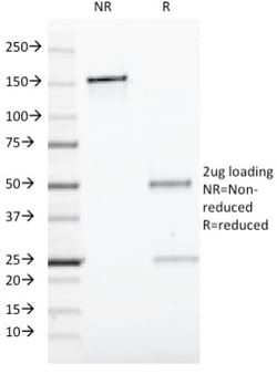
Concentration
0.2mg/mL
Dilution
Flow Cytometry 0.5 - 1 ug/million cells in 0.1 ml, Immunohistochemistry-Paraffin 0.5 - 1.0 ug/ml, Immunofluorescence 1 - 2 ug/ml
Gene Accession No.
P16150
Gene Symbols
SPN
Immunogen
Recombinant human SPN protein
Quantity
0.2 mg
Research Discipline
Immunology
Gene ID (Entrez)
6693
Target Species
Human
Form
Purified
Applications
Flow Cytometry, Immunohistochemistry (Paraffin), Immunofluorescence
Clone
SPN/1094
Conjugate
Unconjugated
Formulation
10mM PBS and 0.05% BSA with 0.05% Sodium Azide
Gene Alias
CD43 antigen, CD43), Galactoglycoprotein, GALGP, Leukocyte sialoglycoprotein, Sialophorin, sialophorin (gpL115, leukosialin, CD43)
Host Species
Mouse
Purification Method
Protein A or G purified
Regulatory Status
RUO
Primary or Secondary
Primary
Test Specificity
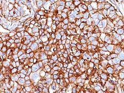
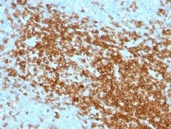
It recognizes a cell surface glycoprotein of 95/115/135kDa (depending upon the extent of glycosylation), identified as CD43. 70-90% of T-cell lymphomas and from 22-37% of B-cell lymphomas express CD43. No reactivity has been observed with reactive B-cells. So a B-lineage population that co-expresses CD43 is highly likely to be a malignant lymphoma, especially a low-grade lymphoma, rather than a reactive B-cell population. When CD43 antibody is used in combination with anti-CD20, effective immunophenotyping of the lymphomas in formalin-fixed tissues can be obtained. Co-staining of a lymphoid infiltrate with anti-CD20 and anti-CD43 argues against a reactive process and favors a diagnosis of lymphoma.
Content And Storage
Store at 4C.
Isotype
IgG1 κ
Related Products


Description
- Ensure accurate, reproducible results in Flow Cytometry, Immunohistochemistry (Paraffin), Immunofluorescence CD43/Sialophorin Monoclonal specifically detects CD43/Sialophorin in Human samples
- It is validated for Western Blot, Flow Cytometry, Immunohistochemistry, Immunocytochemistry/Immunofluorescence, Immunohistochemistry-Paraffin.
Compare Similar Items
Show Difference
Antigen: CD43/Sialophorin
Classification: Monoclonal
Concentration: 0.2mg/mL
Dilution: Flow Cytometry 0.5 - 1 ug/million cells in 0.1 ml, Immunohistochemistry-Paraffin 0.5 - 1.0 ug/ml, Immunofluorescence 1 - 2 ug/ml
Gene Accession No.: P16150
Gene Symbols: SPN
Immunogen: Recombinant human SPN protein
Quantity: 0.2 mg
Research Discipline: Immunology
Gene ID (Entrez): 6693
Target Species: Human
Form: Purified
Applications: Flow Cytometry, Immunohistochemistry (Paraffin), Immunofluorescence
Clone: SPN/1094
Conjugate: Unconjugated
Formulation: 10mM PBS and 0.05% BSA with 0.05% Sodium Azide
Gene Alias: CD43 antigen, CD43), Galactoglycoprotein, GALGP, Leukocyte sialoglycoprotein, Sialophorin, sialophorin (gpL115, leukosialin, CD43)
Host Species: Mouse
Purification Method: Protein A or G purified
Regulatory Status: RUO
Primary or Secondary: Primary
Test Specificity: It recognizes a cell surface glycoprotein of 95/115/135kDa (depending upon the extent of glycosylation), identified as CD43. 70-90% of T-cell lymphomas and from 22-37% of B-cell lymphomas express CD43. No reactivity has been observed with reactive B-cells. So a B-lineage population that co-expresses CD43 is highly likely to be a malignant lymphoma, especially a low-grade lymphoma, rather than a reactive B-cell population. When CD43 antibody is used in combination with anti-CD20, effective immunophenotyping of the lymphomas in formalin-fixed tissues can be obtained. Co-staining of a lymphoid infiltrate with anti-CD20 and anti-CD43 argues against a reactive process and favors a diagnosis of lymphoma.
Content And Storage: Store at 4C.
Isotype: IgG1 κ
Antigen: CD44
Classification: Monoclonal
Concentration: 0.2mg/mL
Dilution: Western Blot, Flow Cytometry 0.5 - 1 ug/million cells in 0.1 ml, Immunohistochemistry-Paraffin 0.25 - 0.5 ug/ml, Immunofluorescence 0.5 - 1.0 ug/ml
Gene Accession No.: __
Gene Symbols: CD44
Immunogen: Recombinant human HCAM protein
Quantity: 0.02 mg
Research Discipline: Cancer, Cell Biology, Extracellular Matrix, Hematopoietic Stem Cell Markers, Immunology, Mesenchymal Stem Cell Markers, Signal Transduction, Stem Cell Markers
Gene ID (Entrez): 960
Target Species: Human, Primate
Form: Purified
Applications: Western Blot, Flow Cytometry, Immunohistochemistry (Paraffin), Immunofluorescence
Clone: HCAM/918
Conjugate: Unconjugated
Formulation: 10mM PBS and 0.05% BSA with 0.05% Sodium Azide
Gene Alias: CD44 antigen, CD44 molecule (Indian blood group), CD44R, CDw44, cell surface glycoprotein CD44, chondroitin sulfate proteoglycan 8, CSPG8, ECMR-III, Epican, Extracellular matrix receptor III, GP90 lymphocyte homing/adhesion receptor, HCELL, hematopoietic cell E- and L-selectin ligand, Heparan sulfate proteoglycan, Hermes antigen, homing function and Indian blood group system, HUTCH-I, Hyaluronate receptor, IN, LHR, MC56, MDU2CD44 antigen (homing function and Indian blood group system), MDU3CDW44, MIC4MGC10468, Pgp1, PGP-1, PGP-I, Phagocytic glycoprotein 1, Phagocytic glycoprotein I
Host Species: Mouse
Purification Method: Protein A or G purified
Regulatory Status: RUO
Primary or Secondary: Primary
Test Specificity: Recognizes a cell surface glycoprotein of 80-95kDa (CD44) on lymphocytes, monocytes, and granulocytes (Leucocyte Typing Workshop V). Its epitope is resistant to digestion by trypsin and chymotrypsin. The CD44 family of glycoproteins exists in a number of variant isoforms, the most common being the standard 85-95kDa or hematopoietic variant (CD44s). Higher molecular weight isoforms are described in epithelial cells (CD44v), which are believed to function in intercellular adhesion and stromal binding. CD44 immunostaining is commonly used for the discrimination of urothelial transitional cell carcinoma in-situ from non-neoplastic changes in the urothelium.
Content And Storage: Store at 4C.
Isotype: IgG2a κ
Antigen: CD44
Classification: Monoclonal
Concentration: 0.2mg/mL
Dilution: Western Blot, Flow Cytometry 0.5 - 1 ug/million cells in 0.1 ml, Immunohistochemistry-Paraffin 0.25 - 0.5 ug/ml, Immunofluorescence 0.5 - 1.0 ug/ml
Gene Accession No.: __
Gene Symbols: CD44
Immunogen: Recombinant human HCAM protein
Quantity: 0.1 mg
Research Discipline: Cancer, Cell Biology, Extracellular Matrix, Hematopoietic Stem Cell Markers, Immunology, Mesenchymal Stem Cell Markers, Signal Transduction, Stem Cell Markers
Gene ID (Entrez): 960
Target Species: Human, Primate
Form: Purified
Applications: Western Blot, Flow Cytometry, Immunohistochemistry (Paraffin), Immunofluorescence
Clone: HCAM/918
Conjugate: Unconjugated
Formulation: 10mM PBS and 0.05% BSA with 0.05% Sodium Azide
Gene Alias: CD44 antigen, CD44 molecule (Indian blood group), CD44R, CDw44, cell surface glycoprotein CD44, chondroitin sulfate proteoglycan 8, CSPG8, ECMR-III, Epican, Extracellular matrix receptor III, GP90 lymphocyte homing/adhesion receptor, HCELL, hematopoietic cell E- and L-selectin ligand, Heparan sulfate proteoglycan, Hermes antigen, homing function and Indian blood group system, HUTCH-I, Hyaluronate receptor, IN, LHR, MC56, MDU2CD44 antigen (homing function and Indian blood group system), MDU3CDW44, MIC4MGC10468, Pgp1, PGP-1, PGP-I, Phagocytic glycoprotein 1, Phagocytic glycoprotein I
Host Species: Mouse
Purification Method: Protein A or G purified
Regulatory Status: RUO
Primary or Secondary: Primary
Test Specificity: Recognizes a cell surface glycoprotein of 80-95kDa (CD44) on lymphocytes, monocytes, and granulocytes (Leucocyte Typing Workshop V). Its epitope is resistant to digestion by trypsin and chymotrypsin. The CD44 family of glycoproteins exists in a number of variant isoforms, the most common being the standard 85-95kDa or hematopoietic variant (CD44s). Higher molecular weight isoforms are described in epithelial cells (CD44v), which are believed to function in intercellular adhesion and stromal binding. CD44 immunostaining is commonly used for the discrimination of urothelial transitional cell carcinoma in-situ from non-neoplastic changes in the urothelium.
Content And Storage: Store at 4C.
Isotype: IgG2a κ