CD19 Antibody (CVID3/429), Novus Biologicals™
Manufacturer: Fischer Scientific
The price for this product is unavailable. Please request a quote
Antigen
CD19
Classification
Monoclonal
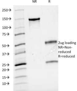
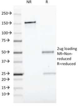
Concentration
0.2 mg/mL
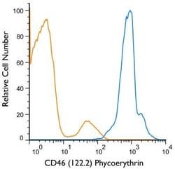
Dilution
Flow Cytometry 0.5 - 1 ug/million cells in 0.1 ml, SDS-Page, Immunofluorescence 0.5 - 1.0 ug/ml
Gene Alias
B4, B-lymphocyte antigen CD19, B-lymphocyte surface antigen B4, CD19 antigen, CD19 molecule, CVID3, Differentiation antigen CD19, MGC12802, T-cell surface antigen Leu-12
Host Species
Mouse
Molecular Weight of Antigen
95 kDa
Quantity
0.2 mg
Research Discipline
Adaptive Immunity, Cytokine Research, Immunology, Innate Immunity, Mesenchymal Stem Cell Markers, Signal Transduction, Stem Cell Lines, Stem Cell Markers
Gene ID (Entrez)
930
Target Species
Human, Monkey, Primate
Form
Purified
Applications
Flow Cytometry, SDS-Page, Immunofluorescence
Clone
CVID3/429
Conjugate
Unconjugated
Formulation
10mM PBS and 0.05% BSA with 0.05% Sodium Azide
Gene Symbols
CD19
Immunogen
Recombinant human CD19 protein
Purification Method
Protein A or G purified
Regulatory Status
RUO
Primary or Secondary
Primary
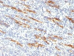
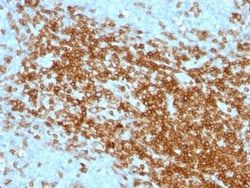
Test Specificity
CD19 is a transmembrane glycoprotein that contains two extracellular immunoglobulin-like domains. CD19 is present in both benign and malignant B-cells and is considered to be the most reliable surface marker of this lineage over a wide range of maturational stages. In normal lymphoid tissue, CD19 is observed in germinal centers, in mantle zone cells, and in scattered cells of the inter-follicular areas. Anti-CD19 exhibits an overall immunoreactivity pattern similar to those of the antibodies against CD20 and CD22. However, in contrast to CD20, expression of CD19 is continuous throughout B-cell development and through terminal differentiation of B-cells into plasma cells. Anti-CD19 positivity is seen in the vast majority of B-cell neoplasms commonly at a lower intensity than normal B-cell counterparts. Plasma cell neoplasms are nearly always negative, as are T-cell neoplasms
Content And Storage
Store at 4C.
Isotype
IgG1 κ
Related Products

Description
- Ensure accurate, reproducible results in Flow Cytometry, Immunofluorescence CD19 Monoclonal specifically detects CD19 in Human, Monkey, Chimpanzee samples
- It is validated for Flow Cytometry, Immunocytochemistry/Immunofluorescence.
Compare Similar Items
Show Difference
Antigen: CD19
Classification: Monoclonal
Concentration: 0.2 mg/mL
Dilution: Flow Cytometry 0.5 - 1 ug/million cells in 0.1 ml, SDS-Page, Immunofluorescence 0.5 - 1.0 ug/ml
Gene Alias: B4, B-lymphocyte antigen CD19, B-lymphocyte surface antigen B4, CD19 antigen, CD19 molecule, CVID3, Differentiation antigen CD19, MGC12802, T-cell surface antigen Leu-12
Host Species: Mouse
Molecular Weight of Antigen: 95 kDa
Quantity: 0.2 mg
Research Discipline: Adaptive Immunity, Cytokine Research, Immunology, Innate Immunity, Mesenchymal Stem Cell Markers, Signal Transduction, Stem Cell Lines, Stem Cell Markers
Gene ID (Entrez): 930
Target Species: Human, Monkey, Primate
Form: Purified
Applications: Flow Cytometry, SDS-Page, Immunofluorescence
Clone: CVID3/429
Conjugate: Unconjugated
Formulation: 10mM PBS and 0.05% BSA with 0.05% Sodium Azide
Gene Symbols: CD19
Immunogen: Recombinant human CD19 protein
Purification Method: Protein A or G purified
Regulatory Status: RUO
Primary or Secondary: Primary
Test Specificity: CD19 is a transmembrane glycoprotein that contains two extracellular immunoglobulin-like domains. CD19 is present in both benign and malignant B-cells and is considered to be the most reliable surface marker of this lineage over a wide range of maturational stages. In normal lymphoid tissue, CD19 is observed in germinal centers, in mantle zone cells, and in scattered cells of the inter-follicular areas. Anti-CD19 exhibits an overall immunoreactivity pattern similar to those of the antibodies against CD20 and CD22. However, in contrast to CD20, expression of CD19 is continuous throughout B-cell development and through terminal differentiation of B-cells into plasma cells. Anti-CD19 positivity is seen in the vast majority of B-cell neoplasms commonly at a lower intensity than normal B-cell counterparts. Plasma cell neoplasms are nearly always negative, as are T-cell neoplasms
Content And Storage: Store at 4C.
Isotype: IgG1 κ

Antigen: Aminopeptidase N/CD13
Classification: Monoclonal
Concentration: 0.2 mg/mL
Dilution: Flow Cytometry 0.5 - 1 ug/million cells in 0.1 ml, Immunohistochemistry-Paraffin 0.5 - 1.0 ug/ml, Immunofluorescence 1 - 2 ug/ml
Gene Alias: alanyl (membrane) aminopeptidase, Alanyl aminopeptidase, Aminopeptidase M, aminopeptidase N, AP-M, AP-N, CD13 antigen, CD13APN, EC 3.4.11, EC 3.4.11.2, gp150, LAP1, Microsomal aminopeptidase, Myeloid plasma membrane glycoprotein CD13, P150, PEPNhAPN
Host Species: Mouse
Molecular Weight of Antigen: 150 kDa
Quantity: 0.02 mg
Research Discipline: Angiogenesis, Cellular Markers, Hematopoietic Stem Cell Markers, Immunology, Mesenchymal Stem Cell Markers, Stem Cell Markers
Gene ID (Entrez): 290
Target Species: Human
Form: Purified
Applications: Flow Cytometry, Immunohistochemistry (Paraffin), Immunofluorescence
Clone: APN/514
Conjugate: Unconjugated
Formulation: 10mM PBS and 0.05% BSA with 0.05% Sodium Azide
Gene Symbols: ANPEP
Immunogen: Recombinant human CD13 protein
Purification Method: Protein A or G purified
Regulatory Status: RUO
Primary or Secondary: Primary
Test Specificity: This MAb recognizes an extracellular epitope of an integral membrane glycoprotein of 150kDa, identified as CD13. This antigen is present on most cells of myeloid origin including granulocytes, monocytes, mast cells, and GM-progenitor cells. It is also expressed by the majority of AML, CML in myeloid blast crisis, and in a smaller fraction of lymphoid leukemias. It is absent from normal lymphocytes, platelets and erythrocytes. CD13 is also present on fibroblasts; endothelial cells, epithelial cells from renal proximal tubules and intestinal brush border, bone marrow stromal cells, osteoclasts, and cells lining bile duct canaliculi. CD13 is identical to aminopeptidase N (APN), a prominent membrane-bound metalloprotease present on the surface of intestinal brush border and renal tubules. CD13 plays a role in metabolism of biologically active peptides, in phagocytosis, and in bactericidal/tumoricidal activities. It also serves as a receptor for human coronaviruses (HCV). The lineage-restricted pattern of expression of CD13 within the hemopoietic compartment suggests that it may be important in myeloid cell differentiation.
Content And Storage: Store at 4C.
Isotype: IgG1 κ
Antigen: Aminopeptidase N/CD13
Classification: Monoclonal
Concentration: 0.2 mg/mL
Dilution: Flow Cytometry 0.5 - 1 ug/million cells in 0.1 ml, Immunohistochemistry-Paraffin 0.5 - 1.0 ug/ml, Immunofluorescence 1 - 2 ug/ml
Gene Alias: alanyl (membrane) aminopeptidase, Alanyl aminopeptidase, Aminopeptidase M, aminopeptidase N, AP-M, AP-N, CD13 antigen, CD13APN, EC 3.4.11, EC 3.4.11.2, gp150, LAP1, Microsomal aminopeptidase, Myeloid plasma membrane glycoprotein CD13, P150, PEPNhAPN
Host Species: Mouse
Molecular Weight of Antigen: 150 kDa
Quantity: 0.1 mg
Research Discipline: Angiogenesis, Cellular Markers, Hematopoietic Stem Cell Markers, Immunology, Mesenchymal Stem Cell Markers, Stem Cell Markers
Gene ID (Entrez): 290
Target Species: Human
Form: Purified
Applications: Flow Cytometry, Immunohistochemistry (Paraffin), Immunofluorescence
Clone: APN/514
Conjugate: Unconjugated
Formulation: 10mM PBS and 0.05% BSA with 0.05% Sodium Azide
Gene Symbols: ANPEP
Immunogen: Recombinant human CD13 protein
Purification Method: Protein A or G purified
Regulatory Status: RUO
Primary or Secondary: Primary
Test Specificity: This MAb recognizes an extracellular epitope of an integral membrane glycoprotein of 150kDa, identified as CD13. This antigen is present on most cells of myeloid origin including granulocytes, monocytes, mast cells, and GM-progenitor cells. It is also expressed by the majority of AML, CML in myeloid blast crisis, and in a smaller fraction of lymphoid leukemias. It is absent from normal lymphocytes, platelets and erythrocytes. CD13 is also present on fibroblasts; endothelial cells, epithelial cells from renal proximal tubules and intestinal brush border, bone marrow stromal cells, osteoclasts, and cells lining bile duct canaliculi. CD13 is identical to aminopeptidase N (APN), a prominent membrane-bound metalloprotease present on the surface of intestinal brush border and renal tubules. CD13 plays a role in metabolism of biologically active peptides, in phagocytosis, and in bactericidal/tumoricidal activities. It also serves as a receptor for human coronaviruses (HCV). The lineage-restricted pattern of expression of CD13 within the hemopoietic compartment suggests that it may be important in myeloid cell differentiation.
Content And Storage: Store at 4C.
Isotype: IgG1 κ