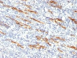
CD99 Antibody (12E7 + MIC2/877), Novus Biologicals™
Manufacturer: Fischer Scientific
Select a Size
| Pack Size | SKU | Availability | Price |
|---|---|---|---|
| Each of 1 | NBP24469701-Each-of-1 | In Stock | ₹ 22,517.00 |
NBP24469701 - Each of 1
In Stock
Quantity
1
Base Price: ₹ 22,517.00
GST (18%): ₹ 4,053.06
Total Price: ₹ 26,570.06
Antigen
CD99
Classification
Monoclonal
Concentration
0.2mg/mL
Dilution
Flow Cytometry 5 - 10 ul/million cells in 0.1ml, Immunohistochemistry-Paraffin 1:100-1:200, Immunofluorescence 1:100-1:200
Gene Accession No.
P14209, P14209
Gene Symbols
CD99
Immunogen
Human acute lymphocytic leukemia T-cells (12E7); Recombinant human MIC2 protein (MIC2/877)
Quantity
0.1 mL
Research Discipline
Immunology
Gene ID (Entrez)
4267
Target Species
Human
Form
Supernatant
Applications
Flow Cytometry, Immunohistochemistry (Paraffin), Immunofluorescence
Clone
12E7 + MIC2/877
Conjugate
Unconjugated
Formulation
No buffer with 0.05% Sodium Azide
Gene Alias
12E7, antigen identified by monoclonal antibodies 12E7, F21 and O13, CD99 antigenY homolog, CD99 molecule, E2 antigen, HBA71, MIC2 (monoclonal 12E7), MIC2Y, MSK5X, Protein MIC2, surface antigen MIC2, T-cell surface glycoprotein E2
Host Species
Mouse
Purification Method
Tissue culture supernatant
Regulatory Status
RUO
Primary or Secondary
Primary
Test Specificity
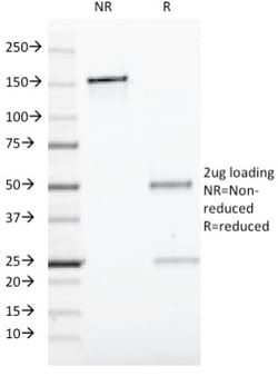
Recognizes a sialoglycoprotein of 27-32kDa, identified as CD99, or MIC2 gene product, or E2 antigen. MIC2 gene is located in the pseudo-autosomal region of the human X and Y chromosome. MIC2 gene encodes two distinct proteins, which are produced by alternative splicing of the CD99 gene transcript and are identified as bands of 30 and 32kDa (p30/32). Although its function is not fully understood, CD99 is implicated in various cellular processes including homotypic aggregation of T cells, upregulation of T cell receptor and MHS molecules, apoptosis of immature thymocytes and leukocyte diapedesis.CD99 is expressed on the cell membrane of some lymphocytes, cortical thymocytes, and granulosa cells of the ovary. Most pancreatic islet cells, Sertoli cells of the testis, and some endothelial cells express this antigen. Mature granulocytes express very little or no CD99. MIC2 is strongly expressed on Ewings sarcoma cells and primitive peripheral neuroectodermal tumors.
Content And Storage
Store at 4C.
Isotype
IgG
Related Products

Description
- Ensure accurate, reproducible results in Flow Cytometry, Immunohistochemistry (Paraffin), Immunofluorescence CD99 Monoclonal specifically detects CD99 in Human samples
- It is validated for Flow Cytometry, Immunohistochemistry, Immunocytochemistry/Immunofluorescence, Immunohistochemistry-Paraffin, Immunofluorescence.
Compare Similar Items
Show Difference
Antigen: CD99
Classification: Monoclonal
Concentration: 0.2mg/mL
Dilution: Flow Cytometry 5 - 10 ul/million cells in 0.1ml, Immunohistochemistry-Paraffin 1:100-1:200, Immunofluorescence 1:100-1:200
Gene Accession No.: P14209, P14209
Gene Symbols: CD99
Immunogen: Human acute lymphocytic leukemia T-cells (12E7); Recombinant human MIC2 protein (MIC2/877)
Quantity: 0.1 mL
Research Discipline: Immunology
Gene ID (Entrez): 4267
Target Species: Human
Form: Supernatant
Applications: Flow Cytometry, Immunohistochemistry (Paraffin), Immunofluorescence
Clone: 12E7 + MIC2/877
Conjugate: Unconjugated
Formulation: No buffer with 0.05% Sodium Azide
Gene Alias: 12E7, antigen identified by monoclonal antibodies 12E7, F21 and O13, CD99 antigenY homolog, CD99 molecule, E2 antigen, HBA71, MIC2 (monoclonal 12E7), MIC2Y, MSK5X, Protein MIC2, surface antigen MIC2, T-cell surface glycoprotein E2
Host Species: Mouse
Purification Method: Tissue culture supernatant
Regulatory Status: RUO
Primary or Secondary: Primary
Test Specificity: Recognizes a sialoglycoprotein of 27-32kDa, identified as CD99, or MIC2 gene product, or E2 antigen. MIC2 gene is located in the pseudo-autosomal region of the human X and Y chromosome. MIC2 gene encodes two distinct proteins, which are produced by alternative splicing of the CD99 gene transcript and are identified as bands of 30 and 32kDa (p30/32). Although its function is not fully understood, CD99 is implicated in various cellular processes including homotypic aggregation of T cells, upregulation of T cell receptor and MHS molecules, apoptosis of immature thymocytes and leukocyte diapedesis.CD99 is expressed on the cell membrane of some lymphocytes, cortical thymocytes, and granulosa cells of the ovary. Most pancreatic islet cells, Sertoli cells of the testis, and some endothelial cells express this antigen. Mature granulocytes express very little or no CD99. MIC2 is strongly expressed on Ewings sarcoma cells and primitive peripheral neuroectodermal tumors.
Content And Storage: Store at 4C.
Isotype: IgG
Antigen: CD99
Classification: Monoclonal
Concentration: 0.2mg/mL
Dilution: Flow Cytometry 5 - 10 ul/million cells in 0.1ml, Immunohistochemistry-Paraffin 1:100-1:200, Immunofluorescence 1:100-1:200
Gene Accession No.: P14209, P14209
Gene Symbols: CD99
Immunogen: Human acute lymphocytic leukemia T-cells (12E7); Recombinant human MIC2 protein (MIC2/877)
Quantity: 0.5 mL
Research Discipline: Immunology
Gene ID (Entrez): 4267
Target Species: Human
Form: Supernatant
Applications: Flow Cytometry, Immunohistochemistry (Paraffin), Immunofluorescence
Clone: 12E7 + MIC2/877
Conjugate: Unconjugated
Formulation: No buffer with 0.05% Sodium Azide
Gene Alias: 12E7, antigen identified by monoclonal antibodies 12E7, F21 and O13, CD99 antigenY homolog, CD99 molecule, E2 antigen, HBA71, MIC2 (monoclonal 12E7), MIC2Y, MSK5X, Protein MIC2, surface antigen MIC2, T-cell surface glycoprotein E2
Host Species: Mouse
Purification Method: Tissue culture supernatant
Regulatory Status: RUO
Primary or Secondary: Primary
Test Specificity: Recognizes a sialoglycoprotein of 27-32kDa, identified as CD99, or MIC2 gene product, or E2 antigen. MIC2 gene is located in the pseudo-autosomal region of the human X and Y chromosome. MIC2 gene encodes two distinct proteins, which are produced by alternative splicing of the CD99 gene transcript and are identified as bands of 30 and 32kDa (p30/32). Although its function is not fully understood, CD99 is implicated in various cellular processes including homotypic aggregation of T cells, upregulation of T cell receptor and MHS molecules, apoptosis of immature thymocytes and leukocyte diapedesis.CD99 is expressed on the cell membrane of some lymphocytes, cortical thymocytes, and granulosa cells of the ovary. Most pancreatic islet cells, Sertoli cells of the testis, and some endothelial cells express this antigen. Mature granulocytes express very little or no CD99. MIC2 is strongly expressed on Ewings sarcoma cells and primitive peripheral neuroectodermal tumors.
Content And Storage: Store at 4C.
Isotype: IgG

Antigen: CD59
Classification: Monoclonal
Concentration: 0.2 mg/mL
Dilution: Flow Cytometry 0.5 - 1 ug/million cells in 0.1 ml, Immunofluorescence 0.5 - 1.0 ug/ml
Gene Accession No.: __
Gene Symbols: CD59
Immunogen: Stimulated human leukocytes
Quantity: 0.02 mg
Research Discipline: Cell Biology, Cellular Markers, Immunology, Signal Transduction, Stem Cell Markers
Gene ID (Entrez): 966
Target Species: Human, Baboon (Negative), Equine (Negative)
Form: Purified
Applications: Flow Cytometry, Immunofluorescence
Clone: 193-27
Conjugate: Unconjugated
Formulation: 10mM PBS and 0.05% BSA with 0.05% Sodium Azide
Gene Alias: 16.3A5, 1F5, 1F5 antigen, 20 kDa homologous restriction factor, CD59 antigen, CD59 antigen p18-20 (antigen identified by monoclonal antibodies 16.3A5, EJ16, CD59 antigen, complement regulatory protein, CD59 glycoprotein, CD59 molecule, complement regulatory protein, EJ16, EJ30, EJ30, EL32 and G344), EL32, FLJ38134, FLJ92039, G344, HRF20, HRF-20, human leukocyte antigen MIC11, Ly-6-like protein, lymphocytic antigen CD59/MEM43, MACIF, MAC-inhibitory protein, MAC-IP, MEM43, MEM43 antigen, membrane attack complex (MAC) inhibition factor, Membrane attack complex inhibition factor, Membrane inhibitor of reactive lysis, MGC2354, MIC11MSK21, MIN1, MIN2, MIN3, MIRL, p18-20, protectin, surface anitgen recognized by monoclonal 16.3A5, T cell-activating protein
Host Species: Mouse
Purification Method: Protein A or G purified
Regulatory Status: RUO
Primary or Secondary: Primary
Test Specificity: Reacts with human CD59, a 20kDa glycosyl phosphatidyl-inositol (GPI)-anchored cell surface protein (Workshop VI; Code N-L036). CD59 regulates complement-mediated cell lysis, and it is involved in lymphocyte signal transduction. This protein is a potent inhibitor of the complement membrane attack complex, whereby it binds complement C8 and/or C9 during the assembly of this complex, thereby inhibiting the incorporation of multiple copies of C9 into the complex, which is necessary for osmolytic pore formation. CD59 is widely distributed on cells in all tissues. It inhibits formation of MAC, thus protecting cells from complement-mediated lysis. The expression of CD59 on erythrocytes is important for their survival. Genetic defects in GPI-anchor attachment, that cause a reduction or loss of CD59 and CD55 on erythrocytes produce the symptoms of the disease paroxysmal hemoglobinuria (PNH). This MAb recognizes CD59 transfected cells. It is useful for study on GPI-anchored proteins, PNH and CD59 functions.
Content And Storage: Store at 4C.
Isotype: IgM κ