CD59 Antibody (193-27), Novus Biologicals™
Manufacturer: Fischer Scientific
Select a Size
| Pack Size | SKU | Availability | Price |
|---|---|---|---|
| Each of 1 | NBP24469801-Each-of-1 | In Stock | ₹ 46,636.00 |
NBP24469801 - Each of 1
In Stock
Quantity
1
Base Price: ₹ 46,636.00
GST (18%): ₹ 8,394.48
Total Price: ₹ 55,030.48
Antigen
CD59
Classification
Monoclonal
Concentration
0.2 mg/mL
Dilution
Flow Cytometry 0.5 - 1 ug/million cells in 0.1 ml, Immunofluorescence 0.5 - 1.0 ug/ml
Gene Alias
16.3A5, 1F5, 1F5 antigen, 20 kDa homologous restriction factor, CD59 antigen, CD59 antigen p18-20 (antigen identified by monoclonal antibodies 16.3A5, EJ16, CD59 antigen, complement regulatory protein, CD59 glycoprotein, CD59 molecule, complement regulatory protein, EJ16, EJ30, EJ30, EL32 and G344), EL32, FLJ38134, FLJ92039, G344, HRF20, HRF-20, human leukocyte antigen MIC11, Ly-6-like protein, lymphocytic antigen CD59/MEM43, MACIF, MAC-inhibitory protein, MAC-IP, MEM43, MEM43 antigen, membrane attack complex (MAC) inhibition factor, Membrane attack complex inhibition factor, Membrane inhibitor of reactive lysis, MGC2354, MIC11MSK21, MIN1, MIN2, MIN3, MIRL, p18-20, protectin, surface anitgen recognized by monoclonal 16.3A5, T cell-activating protein
Host Species
Mouse
Molecular Weight of Antigen
20 kDa
Quantity
0.1 mg
Research Discipline
Cell Biology, Cellular Markers, Immunology, Signal Transduction, Stem Cell Markers
Gene ID (Entrez)
966
Target Species
Human, Baboon (Negative), Equine (Negative)
Form
Purified
Applications
Flow Cytometry, Immunofluorescence
Clone
193-27
Conjugate
Unconjugated
Formulation
10mM PBS and 0.05% BSA with 0.05% Sodium Azide
Gene Symbols
CD59
Immunogen
Stimulated human leukocytes
Purification Method
Protein A or G purified
Regulatory Status
RUO
Primary or Secondary
Primary
Test Specificity
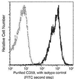
Reacts with human CD59, a 20kDa glycosyl phosphatidyl-inositol (GPI)-anchored cell surface protein (Workshop VI; Code N-L036). CD59 regulates complement-mediated cell lysis, and it is involved in lymphocyte signal transduction. This protein is a potent inhibitor of the complement membrane attack complex, whereby it binds complement C8 and/or C9 during the assembly of this complex, thereby inhibiting the incorporation of multiple copies of C9 into the complex, which is necessary for osmolytic pore formation. CD59 is widely distributed on cells in all tissues. It inhibits formation of MAC, thus protecting cells from complement-mediated lysis. The expression of CD59 on erythrocytes is important for their survival. Genetic defects in GPI-anchor attachment, that cause a reduction or loss of CD59 and CD55 on erythrocytes produce the symptoms of the disease paroxysmal hemoglobinuria (PNH). This MAb recognizes CD59 transfected cells. It is useful for study on GPI-anchored proteins, PNH and CD59 functions.
Content And Storage
Store at 4C.
Isotype
IgM κ
Related Products


Description
- Ensure accurate, reproducible results in Flow Cytometry, Immunofluorescence CD59 Monoclonal specifically detects CD59 in Human, Baboon (Negative), Equine (Negative) samples
- It is validated for Flow Cytometry, Immunocytochemistry/Immunofluorescence, Immunohistochemistry-Paraffin, Functional, Immunofluorescence.
Compare Similar Items
Show Difference
Antigen: CD59
Classification: Monoclonal
Concentration: 0.2 mg/mL
Dilution: Flow Cytometry 0.5 - 1 ug/million cells in 0.1 ml, Immunofluorescence 0.5 - 1.0 ug/ml
Gene Alias: 16.3A5, 1F5, 1F5 antigen, 20 kDa homologous restriction factor, CD59 antigen, CD59 antigen p18-20 (antigen identified by monoclonal antibodies 16.3A5, EJ16, CD59 antigen, complement regulatory protein, CD59 glycoprotein, CD59 molecule, complement regulatory protein, EJ16, EJ30, EJ30, EL32 and G344), EL32, FLJ38134, FLJ92039, G344, HRF20, HRF-20, human leukocyte antigen MIC11, Ly-6-like protein, lymphocytic antigen CD59/MEM43, MACIF, MAC-inhibitory protein, MAC-IP, MEM43, MEM43 antigen, membrane attack complex (MAC) inhibition factor, Membrane attack complex inhibition factor, Membrane inhibitor of reactive lysis, MGC2354, MIC11MSK21, MIN1, MIN2, MIN3, MIRL, p18-20, protectin, surface anitgen recognized by monoclonal 16.3A5, T cell-activating protein
Host Species: Mouse
Molecular Weight of Antigen: 20 kDa
Quantity: 0.1 mg
Research Discipline: Cell Biology, Cellular Markers, Immunology, Signal Transduction, Stem Cell Markers
Gene ID (Entrez): 966
Target Species: Human, Baboon (Negative), Equine (Negative)
Form: Purified
Applications: Flow Cytometry, Immunofluorescence
Clone: 193-27
Conjugate: Unconjugated
Formulation: 10mM PBS and 0.05% BSA with 0.05% Sodium Azide
Gene Symbols: CD59
Immunogen: Stimulated human leukocytes
Purification Method: Protein A or G purified
Regulatory Status: RUO
Primary or Secondary: Primary
Test Specificity: Reacts with human CD59, a 20kDa glycosyl phosphatidyl-inositol (GPI)-anchored cell surface protein (Workshop VI; Code N-L036). CD59 regulates complement-mediated cell lysis, and it is involved in lymphocyte signal transduction. This protein is a potent inhibitor of the complement membrane attack complex, whereby it binds complement C8 and/or C9 during the assembly of this complex, thereby inhibiting the incorporation of multiple copies of C9 into the complex, which is necessary for osmolytic pore formation. CD59 is widely distributed on cells in all tissues. It inhibits formation of MAC, thus protecting cells from complement-mediated lysis. The expression of CD59 on erythrocytes is important for their survival. Genetic defects in GPI-anchor attachment, that cause a reduction or loss of CD59 and CD55 on erythrocytes produce the symptoms of the disease paroxysmal hemoglobinuria (PNH). This MAb recognizes CD59 transfected cells. It is useful for study on GPI-anchored proteins, PNH and CD59 functions.
Content And Storage: Store at 4C.
Isotype: IgM κ
Antigen: CD59
Classification: Monoclonal
Concentration: 0.2 mg/mL
Dilution: Flow Cytometry 0.5 - 1 ug/million cells in 0.1 ml, Immunofluorescence 0.5 - 1.0 ug/ml
Gene Alias: 16.3A5, 1F5, 1F5 antigen, 20 kDa homologous restriction factor, CD59 antigen, CD59 antigen p18-20 (antigen identified by monoclonal antibodies 16.3A5, EJ16, CD59 antigen, complement regulatory protein, CD59 glycoprotein, CD59 molecule, complement regulatory protein, EJ16, EJ30, EJ30, EL32 and G344), EL32, FLJ38134, FLJ92039, G344, HRF20, HRF-20, human leukocyte antigen MIC11, Ly-6-like protein, lymphocytic antigen CD59/MEM43, MACIF, MAC-inhibitory protein, MAC-IP, MEM43, MEM43 antigen, membrane attack complex (MAC) inhibition factor, Membrane attack complex inhibition factor, Membrane inhibitor of reactive lysis, MGC2354, MIC11MSK21, MIN1, MIN2, MIN3, MIRL, p18-20, protectin, surface anitgen recognized by monoclonal 16.3A5, T cell-activating protein
Host Species: Mouse
Molecular Weight of Antigen: 20 kDa
Quantity: 0.2 mg
Research Discipline: Cell Biology, Cellular Markers, Immunology, Signal Transduction, Stem Cell Markers
Gene ID (Entrez): 966
Target Species: Human, Baboon (Negative), Equine (Negative)
Form: Purified
Applications: Flow Cytometry, Immunofluorescence
Clone: 193-27
Conjugate: Unconjugated
Formulation: 10mM PBS and 0.05% BSA with 0.05% Sodium Azide
Gene Symbols: CD59
Immunogen: Stimulated human leukocytes
Purification Method: Protein A or G purified
Regulatory Status: RUO
Primary or Secondary: Primary
Test Specificity: Reacts with human CD59, a 20kDa glycosyl phosphatidyl-inositol (GPI)-anchored cell surface protein (Workshop VI; Code N-L036). CD59 regulates complement-mediated cell lysis, and it is involved in lymphocyte signal transduction. This protein is a potent inhibitor of the complement membrane attack complex, whereby it binds complement C8 and/or C9 during the assembly of this complex, thereby inhibiting the incorporation of multiple copies of C9 into the complex, which is necessary for osmolytic pore formation. CD59 is widely distributed on cells in all tissues. It inhibits formation of MAC, thus protecting cells from complement-mediated lysis. The expression of CD59 on erythrocytes is important for their survival. Genetic defects in GPI-anchor attachment, that cause a reduction or loss of CD59 and CD55 on erythrocytes produce the symptoms of the disease paroxysmal hemoglobinuria (PNH). This MAb recognizes CD59 transfected cells. It is useful for study on GPI-anchored proteins, PNH and CD59 functions.
Content And Storage: Store at 4C.
Isotype: IgM κ
Antigen: CD59
Classification: Monoclonal
Concentration: 0.2 mg/mL
Dilution: Flow Cytometry 0.5 - 1 ug/million cells in 0.1 ml, SDS-Page, Immunofluorescence 0.5 - 1.0 ug/ml
Gene Alias: 16.3A5, 1F5, 1F5 antigen, 20 kDa homologous restriction factor, CD59 antigen, CD59 antigen p18-20 (antigen identified by monoclonal antibodies 16.3A5, EJ16, CD59 antigen, complement regulatory protein, CD59 glycoprotein, CD59 molecule, complement regulatory protein, EJ16, EJ30, EJ30, EL32 and G344), EL32, FLJ38134, FLJ92039, G344, HRF20, HRF-20, human leukocyte antigen MIC11, Ly-6-like protein, lymphocytic antigen CD59/MEM43, MACIF, MAC-inhibitory protein, MAC-IP, MEM43, MEM43 antigen, membrane attack complex (MAC) inhibition factor, Membrane attack complex inhibition factor, Membrane inhibitor of reactive lysis, MGC2354, MIC11MSK21, MIN1, MIN2, MIN3, MIRL, p18-20, protectin, surface anitgen recognized by monoclonal 16.3A5, T cell-activating protein
Host Species: Mouse
Molecular Weight of Antigen: 20 kDa
Quantity: 0.02 mg
Research Discipline: Cell Biology, Cellular Markers, Immunology, Signal Transduction, Stem Cell Markers
Gene ID (Entrez): 966
Target Species: Human
Form: Purified
Applications: Flow Cytometry, SDS-Page, Immunofluorescence
Clone: BRA-10G
Conjugate: Unconjugated
Formulation: 10mM PBS and 0.05% BSA with 0.05% Sodium Azide
Gene Symbols: CD59
Immunogen: Human K562 tumor cells
Purification Method: Protein A or G purified
Regulatory Status: RUO
Primary or Secondary: Primary
Test Specificity: Reacts with human CD59, a 20kDa glycosyl phosphatidyl-inositol (GPI)-anchored cell surface protein. CD59 regulates complement-mediated cell lysis, and it is involved in lymphocyte signal transduction. This protein is a potent inhibitor of the complement membrane attack complex, whereby it binds complement C8 and/or C9 during the assembly of this complex, thereby inhibiting the incorporation of multiple copies of C9 into the complex, which is necessary for osmolytic pore formation. CD59 is widely distributed on cells in all tissues. It inhibits formation of MAC, thus protecting cells from complement-mediated lysis. The expression of CD59 on erythrocytes is important for their survival. Genetic defects in GPI-anchor attachment, that cause a reduction or loss of CD59 and CD55 on erythrocytes produce the symptoms of the disease paroxysmal hemoglobinuria (PNH). This MAb is useful for study on GPI-anchored proteins, PNH and CD59 functions.
Content And Storage: Store at 4C.
Isotype: IgG1 κ