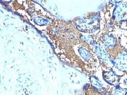
Glycophorin A Antibody (SPM599), Novus Biologicals™
Manufacturer: Fischer Scientific
The price for this product is unavailable. Please request a quote
Antigen
Glycophorin A
Classification
Monoclonal
Concentration
0.2mg/mL
Dilution
Western Blot, Flow Cytometry 0.5 - 1 ug/million cells in 0.1 ml, Immunohistochemistry-Paraffin 0.25 - 0.5 ug/ml, Immunofluorescence 0.5 - 1.0 ug/ml
Gene Accession No.
P02724
Gene Symbols
GYPA
Immunogen
Recombinant human glycophorin A protein
Purification Method
Protein A or G purified
Regulatory Status
RUO
Primary or Secondary
Primary
Test Specificity
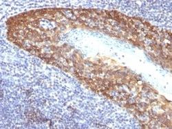
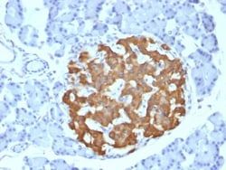
Recognizes a sialoglycoprotein of 39kDa, identified as glycophorin A (GPA). It is present on red blood cells (RBC) and erythroid precursor cells. It has been shown that glycophorin acts as the receptor for Sandei virus and parvovirus. Glycophorins A (GPA) and B (GPB), which are single, trans-membrane sialoglycoproteins. GPA is the carrier of blood group M and N specificities, while GPB accounts for S and U specificities. GPA and GPB provide the cells with a large mucin like surface and it has been suggested this provides a barrier to cell fusion, so minimizing aggregation between red blood cells in the circulation.
Content And Storage
Store at 4C.
Isotype
IgG1 κ
Applications
Western Blot, Flow Cytometry, Immunohistochemistry (Paraffin), Immunofluorescence
Clone
SPM599
Conjugate
Unconjugated
Formulation
10mM PBS and 0.05% BSA with 0.05% Sodium Azide
Gene Alias
CD235a antigen, glycophorin A (includes MN blood group), glycophorin A (MNS blood group), glycophorin Erik, glycophorin MiI, glycophorin MiIII, glycophorin MiV, glycophorin MiX, glycophorin SAT, glycophorin Sta type C, glycophorin-A, GPA, GPErik, GpMiIII, GPSAT, HGpMiIII, HGpMiV, HGpMiX, HGpMiXI, HGpSta(C), Mi.V glycoprotein (24 AA), MN sialoglycoprotein, MNS, PAS-2, recombinant glycophorin A-B Miltenberger-DR
Host Species
Mouse
Molecular Weight of Antigen
39 kDa
Quantity
0.2 mg
Research Discipline
Cancer, Signal Transduction
Gene ID (Entrez)
2993
Target Species
Human
Form
Purified
Related Products

Description
- Ensure accurate, reproducible results in Western Blot, Flow Cytometry, Immunohistochemistry (Paraffin), Immunofluorescence Glycophorin A Monoclonal specifically detects Glycophorin A in Human samples
- It is validated for Western Blot, Immunohistochemistry, Immunohistochemistry-Paraffin.
Compare Similar Items
Show Difference
Antigen: Glycophorin A
Classification: Monoclonal
Concentration: 0.2mg/mL
Dilution: Western Blot, Flow Cytometry 0.5 - 1 ug/million cells in 0.1 ml, Immunohistochemistry-Paraffin 0.25 - 0.5 ug/ml, Immunofluorescence 0.5 - 1.0 ug/ml
Gene Accession No.: P02724
Gene Symbols: GYPA
Immunogen: Recombinant human glycophorin A protein
Purification Method: Protein A or G purified
Regulatory Status: RUO
Primary or Secondary: Primary
Test Specificity: Recognizes a sialoglycoprotein of 39kDa, identified as glycophorin A (GPA). It is present on red blood cells (RBC) and erythroid precursor cells. It has been shown that glycophorin acts as the receptor for Sandei virus and parvovirus. Glycophorins A (GPA) and B (GPB), which are single, trans-membrane sialoglycoproteins. GPA is the carrier of blood group M and N specificities, while GPB accounts for S and U specificities. GPA and GPB provide the cells with a large mucin like surface and it has been suggested this provides a barrier to cell fusion, so minimizing aggregation between red blood cells in the circulation.
Content And Storage: Store at 4C.
Isotype: IgG1 κ
Applications: Western Blot, Flow Cytometry, Immunohistochemistry (Paraffin), Immunofluorescence
Clone: SPM599
Conjugate: Unconjugated
Formulation: 10mM PBS and 0.05% BSA with 0.05% Sodium Azide
Gene Alias: CD235a antigen, glycophorin A (includes MN blood group), glycophorin A (MNS blood group), glycophorin Erik, glycophorin MiI, glycophorin MiIII, glycophorin MiV, glycophorin MiX, glycophorin SAT, glycophorin Sta type C, glycophorin-A, GPA, GPErik, GpMiIII, GPSAT, HGpMiIII, HGpMiV, HGpMiX, HGpMiXI, HGpSta(C), Mi.V glycoprotein (24 AA), MN sialoglycoprotein, MNS, PAS-2, recombinant glycophorin A-B Miltenberger-DR
Host Species: Mouse
Molecular Weight of Antigen: 39 kDa
Quantity: 0.2 mg
Research Discipline: Cancer, Signal Transduction
Gene ID (Entrez): 2993
Target Species: Human
Form: Purified
Antigen: Cytokeratin 14
Classification: Monoclonal
Concentration: 0.2mg/mL
Dilution: Flow Cytometry 0.5 - 1 ug/million cells in 0.1 ml, Immunohistochemistry-Paraffin 0.5 - 1.0 ug/ml, Immunofluorescence 0.5 - 1.0 ug/ml
Gene Accession No.: P02533
Gene Symbols: KRT14
Immunogen: Recombinant full-length human KRT14 protein
Purification Method: Protein A or G purified
Regulatory Status: RUO
Primary or Secondary: Primary
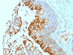
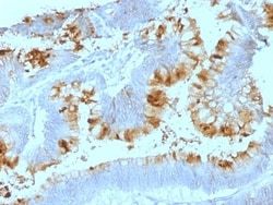
Test Specificity: Cytokeratin 14 (CK14) belongs to the type I (or A or acidic) subfamily of low molecular weight keratins and exists in combination with keratin 5 (type II or B or basic). CK14 is found in basal cells of squamous epithelia, some glandular epithelia, myoepithelium, and mesothelial cells. Anti-CK14 is useful in differentiating squamous cell carcinomas from poorly differentiated epithelial tumors. Anti-CK14 is one of the specific basal markers for distinguishing between basal and non-basal subtypes of breast carcinomas. Anti-CK14 is also a good marker for differentiation of intraductal from invasive salivary duct carcinoma by the positive staining of basal cells surrounding the in-situ neoplasm as well as for differentiation of benign prostate from prostate carcinoma. Furthermore, this antibody has been useful in separating oncocytic tumors of the kidney from its renal mimics, and in identifying metaplastic carcinomas of the breast.
Content And Storage: Store at 4C.
Isotype: IgG3
Applications: Flow Cytometry, Immunohistochemistry (Paraffin), Immunofluorescence
Clone: KRT14/532
Conjugate: Unconjugated
Formulation: 10mM PBS and 0.05% BSA with 0.05% Sodium Azide
Gene Alias: CK14, CK-14, cytokeratin 14, cytokeratin-14, EBS3, EBS4, K14, keratin 14, keratin 14 (epidermolysis bullosa simplex, Dowling-Meara, Koebner), keratin, type I cytoskeletal 14, Keratin-14, NFJ
Host Species: Mouse
Molecular Weight of Antigen: 50 kDa
Quantity: 0.02 mg
Research Discipline: Cytoskeleton Markers
Gene ID (Entrez): 3861
Target Species: Human, Mouse, Rat
Form: Purified
Antigen: Cytokeratin 14
Classification: Monoclonal
Concentration: 0.2mg/mL
Dilution: Flow Cytometry 0.5 - 1 ug/million cells in 0.1 ml, Immunohistochemistry-Paraffin 0.5 - 1.0 ug/ml, Immunofluorescence 0.5 - 1.0 ug/ml
Gene Accession No.: P02533
Gene Symbols: KRT14
Immunogen: Recombinant full-length human KRT14 protein
Purification Method: Protein A or G purified
Regulatory Status: RUO
Primary or Secondary: Primary
Test Specificity: Cytokeratin 14 (CK14) belongs to the type I (or A or acidic) subfamily of low molecular weight keratins and exists in combination with keratin 5 (type II or B or basic). CK14 is found in basal cells of squamous epithelia, some glandular epithelia, myoepithelium, and mesothelial cells. Anti-CK14 is useful in differentiating squamous cell carcinomas from poorly differentiated epithelial tumors. Anti-CK14 is one of the specific basal markers for distinguishing between basal and non-basal subtypes of breast carcinomas. Anti-CK14 is also a good marker for differentiation of intraductal from invasive salivary duct carcinoma by the positive staining of basal cells surrounding the in-situ neoplasm as well as for differentiation of benign prostate from prostate carcinoma. Furthermore, this antibody has been useful in separating oncocytic tumors of the kidney from its renal mimics, and in identifying metaplastic carcinomas of the breast.
Content And Storage: Store at 4C.
Isotype: IgG3
Applications: Flow Cytometry, Immunohistochemistry (Paraffin), Immunofluorescence
Clone: KRT14/532
Conjugate: Unconjugated
Formulation: 10mM PBS and 0.05% BSA with 0.05% Sodium Azide
Gene Alias: CK14, CK-14, cytokeratin 14, cytokeratin-14, EBS3, EBS4, K14, keratin 14, keratin 14 (epidermolysis bullosa simplex, Dowling-Meara, Koebner), keratin, type I cytoskeletal 14, Keratin-14, NFJ
Host Species: Mouse
Molecular Weight of Antigen: 50 kDa
Quantity: 0.1 mg
Research Discipline: Cytoskeleton Markers
Gene ID (Entrez): 3861
Target Species: Human, Mouse, Rat
Form: Purified