Hepatocyte Specific Antigen Antibody (HepPar1), Novus Biologicals™
Manufacturer: Fischer Scientific
Select a Size
| Pack Size | SKU | Availability | Price |
|---|---|---|---|
| Each of 1 | NBP24527201-Each-of-1 | In Stock | ₹ 45,568.00 |
NBP24527201 - Each of 1
In Stock
Quantity
1
Base Price: ₹ 45,568.00
GST (18%): ₹ 8,202.24
Total Price: ₹ 53,770.24
Antigen
Hepatocyte Specific Antigen
Classification
Monoclonal
Concentration
0.2mg/mL
Dilution
Immunohistochemistry, Immunohistochemistry-Paraffin 0.25 - 0.5 ug/ml, Immunofluorescence 0.5 - 1.0 ug/ml
Gene Alias
Hepatocyte, HSA
Immunogen
Extract of a formalin-fixed, rejected-allograft of a human liver
Quantity
0.1 mg
Primary or Secondary
Primary
Target Species
Human, Mouse, Canine
Form
Purified
Applications
Immunohistochemistry, Immunohistochemistry (Paraffin), Immunofluorescence
Clone
HepPar1
Conjugate
Unconjugated
Formulation
10mM PBS and 0.05% BSA with 0.05% Sodium Azide
Host Species
Mouse
Purification Method
Protein A or G purified
Regulatory Status
RUO
Test Specificity
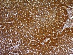
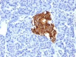
Hepatocyte Paraffin 1 or HepPar1 localizes to the mitochondria of hepatocytes. It is a sensitive marker for distinguishing hepatocellular carcinomas (HCC) from other metastatic carcinomas as well as cholangio-carcinomas. HCC s occur primarily in the stomach, but they are also found in many other organs. The Hepatocyte Specific Antigen may also be a useful marker for intestinal metaplasia. Reportedly, strong expression of the Hepatocyte Specific Antigen correlates with smaller tumor size and longer patient survival. Occasionally, Hepatocyte Specific Antigen is also found in gastric carcinomas as well as in a few other non-hepatic tumors.
Content And Storage
Store at 4C.
Isotype
IgG1
Related Products

Description
- Ensure accurate, reproducible results in Immunohistochemistry (Paraffin), Immunofluorescence Hepatocyte Specific Antigen Monoclonal specifically detects Hepatocyte Specific Antigen in Human, Mouse, Canine samples
- It is validated for Western Blot, Immunohistochemistry, Immunocytochemistry/Immunofluorescence, Immunohistochemistry-Paraffin, Immunofluorescence.
Compare Similar Items
Show Difference
Antigen: Hepatocyte Specific Antigen
Classification: Monoclonal
Concentration: 0.2mg/mL
Dilution: Immunohistochemistry, Immunohistochemistry-Paraffin 0.25 - 0.5 ug/ml, Immunofluorescence 0.5 - 1.0 ug/ml
Gene Alias: Hepatocyte, HSA
Immunogen: Extract of a formalin-fixed, rejected-allograft of a human liver
Quantity: 0.1 mg
Primary or Secondary: Primary
Target Species: Human, Mouse, Canine
Form: Purified
Applications: Immunohistochemistry, Immunohistochemistry (Paraffin), Immunofluorescence
Clone: HepPar1
Conjugate: Unconjugated
Formulation: 10mM PBS and 0.05% BSA with 0.05% Sodium Azide
Host Species: Mouse
Purification Method: Protein A or G purified
Regulatory Status: RUO
Test Specificity: Hepatocyte Paraffin 1 or HepPar1 localizes to the mitochondria of hepatocytes. It is a sensitive marker for distinguishing hepatocellular carcinomas (HCC) from other metastatic carcinomas as well as cholangio-carcinomas. HCC s occur primarily in the stomach, but they are also found in many other organs. The Hepatocyte Specific Antigen may also be a useful marker for intestinal metaplasia. Reportedly, strong expression of the Hepatocyte Specific Antigen correlates with smaller tumor size and longer patient survival. Occasionally, Hepatocyte Specific Antigen is also found in gastric carcinomas as well as in a few other non-hepatic tumors.
Content And Storage: Store at 4C.
Isotype: IgG1
Antigen: Hepatocyte Specific Antigen
Classification: Monoclonal
Concentration: 0.2mg/mL
Dilution: Immunohistochemistry, Immunohistochemistry-Paraffin 0.25 - 0.5 ug/ml, Immunofluorescence 0.5 - 1.0 ug/ml
Gene Alias: Hepatocyte, HSA
Immunogen: Extract of a formalin-fixed, rejected-allograft of a human liver
Quantity: 0.2 mg
Primary or Secondary: Primary
Target Species: Human, Mouse, Canine
Form: Purified
Applications: Immunohistochemistry, Immunohistochemistry (Paraffin), Immunofluorescence
Clone: HepPar1
Conjugate: Unconjugated
Formulation: 10mM PBS and 0.05% BSA with 0.05% Sodium Azide
Host Species: Mouse
Purification Method: Protein A or G purified
Regulatory Status: RUO
Test Specificity: Hepatocyte Paraffin 1 or HepPar1 localizes to the mitochondria of hepatocytes. It is a sensitive marker for distinguishing hepatocellular carcinomas (HCC) from other metastatic carcinomas as well as cholangio-carcinomas. HCC s occur primarily in the stomach, but they are also found in many other organs. The Hepatocyte Specific Antigen may also be a useful marker for intestinal metaplasia. Reportedly, strong expression of the Hepatocyte Specific Antigen correlates with smaller tumor size and longer patient survival. Occasionally, Hepatocyte Specific Antigen is also found in gastric carcinomas as well as in a few other non-hepatic tumors.
Content And Storage: Store at 4C.
Isotype: IgG1
Antigen: pan Actin
Classification: Monoclonal
Concentration: 0.2mg/mL
Dilution: Flow Cytometry 0.5 - 1 ug/million cells in 0.1 ml, Immunohistochemistry-Paraffin 0.25 - 0.5 ug/ml, Immunofluorescence 0.5 - 1.0 ug/ml
Gene Alias: ACTA, actin, alpha 1, skeletal muscle, alpha skeletal muscle, alpha skeletal muscle actin, alpha-actin-1, ASMA, CFTD, CFTDM, MPFD, NEM1, NEM2, NEM3
Immunogen: Recombinant human actin fragment
Quantity: 0.02 mg
Primary or Secondary: Primary
Target Species: Human, Rat, Rabbit
Form: Purified
Applications: Flow Cytometry, Immunohistochemistry (Paraffin), Immunofluorescence
Clone: HHF35 + MSA/953
Conjugate: Unconjugated
Formulation: 10mM PBS and 0.05% BSA with 0.05% Sodium Azide
Host Species: Mouse
Purification Method: Protein A or G purified
Regulatory Status: RUO
Test Specificity: This antibody recognizes actin of skeletal, cardiac, and smooth muscle cells. It is not reactive with other mesenchymal cells except for myoepithelium. Actin can be resolved on the basis of its isoelectric points into three distinctive components: alpha, beta, and gamma in order of increasing isoelectric point. Anti-muscle specific actin recognizes alpha and gamma isotypes of all muscle groups. Non-muscle cells such as vascular endothelial cells and connective tissues are non-reactive. Also, neoplastic cells of non-muscle-derived tissue such as carcinomas, melanomas, and lymphomas are negative.AIt stains tumors of smooth muscle (leiomyomas and leiomyosarcomas) as well as skeletal muscle (rhabdomyomas and rhabdomyosarcomas).
Content And Storage: Store at 4C.
Isotype: IgG1 κ