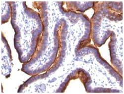
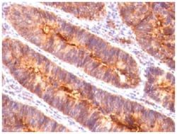
MCAM/CD146 Antibody (MUC18/1130) - Azide and BSA Free, Novus Biologicals™
Manufacturer: Fischer Scientific
The price for this product is unavailable. Please request a quote
Antigen
MCAM/CD146
Classification
Monoclonal
Concentration
1.0 mg/mL
Dilution
Flow Cytometry : 0.5 - 1 ug/million cells in 0.1 ml, Immunohistochemistry-Paraffin : 0.5 - 1.0 ug/ml, Immunofluorescence : 0.5 - 1.0 ug/ml, CyTOF-ready
Gene Alias
CD146, CD146 antigen, cell surface glycoprotein MUC18, Cell surface glycoprotein P1H12, melanoma adhesion molecule, melanoma cell adhesion moleculeS-endo 1 endothelial-associated antigen, Melanoma-associated antigen A32, Melanoma-associated antigen MUC18, MUC18Gicerin
Host Species
Mouse
Molecular Weight of Antigen
130 kDa
Quantity
0.2 mg
Research Discipline
Cancer, Mesenchymal Stem Cell Markers, Stem Cell Markers
Gene ID (Entrez)
4162
Target Species
Human
Form
Purified
Applications
Flow Cytometry, Immunohistochemistry (Paraffin), Immunofluorescence, CyTOF
Clone
MUC18/1130
Conjugate
Unconjugated
Formulation
PBS with No Preservative
Gene Symbols
MCAM
Immunogen
Recombinant human MUC18 protein
Purification Method
Protein A or G purified
Regulatory Status
RUO
Primary or Secondary
Primary
Test Specificity
The human Mel-CAM gene maps to chromosome 11q23 and encodes a trans-membrane glycoprotein, also designated MCAM, MUC 18 or CD146, that belongs to the immunoglobulin superfamily and functions as a Ca2+-independent cell adhesion molecule. Mel-CAM expression is restricted to advanced primary and metastatic melanomas and to cell lines of the neuroectodermal lineage, but not normal melanocytes. Mel-CAM is found on 80% of advanced primary human melanomas and correlates well with development of metastatic disease.
Content And Storage
Store at 4C short term. Aliquot and store at -20C long term. Avoid freeze-thaw cycles.
Isotype
IgG1 κ
Related Products

Description
- MCAM/CD146 Monoclonal specifically detects MCAM/CD146 in Human samples
- It is validated for Flow Cytometry, Immunohistochemistry, Immunocytochemistry/Immunofluorescence, Immunohistochemistry-Paraffin, Immunofluorescence, CyTOF-ready.
Compare Similar Items
Show Difference
Antigen: MCAM/CD146
Classification: Monoclonal
Concentration: 1.0 mg/mL
Dilution: Flow Cytometry : 0.5 - 1 ug/million cells in 0.1 ml, Immunohistochemistry-Paraffin : 0.5 - 1.0 ug/ml, Immunofluorescence : 0.5 - 1.0 ug/ml, CyTOF-ready
Gene Alias: CD146, CD146 antigen, cell surface glycoprotein MUC18, Cell surface glycoprotein P1H12, melanoma adhesion molecule, melanoma cell adhesion moleculeS-endo 1 endothelial-associated antigen, Melanoma-associated antigen A32, Melanoma-associated antigen MUC18, MUC18Gicerin
Host Species: Mouse
Molecular Weight of Antigen: 130 kDa
Quantity: 0.2 mg
Research Discipline: Cancer, Mesenchymal Stem Cell Markers, Stem Cell Markers
Gene ID (Entrez): 4162
Target Species: Human
Form: Purified
Applications: Flow Cytometry, Immunohistochemistry (Paraffin), Immunofluorescence, CyTOF
Clone: MUC18/1130
Conjugate: Unconjugated
Formulation: PBS with No Preservative
Gene Symbols: MCAM
Immunogen: Recombinant human MUC18 protein
Purification Method: Protein A or G purified
Regulatory Status: RUO
Primary or Secondary: Primary
Test Specificity: The human Mel-CAM gene maps to chromosome 11q23 and encodes a trans-membrane glycoprotein, also designated MCAM, MUC 18 or CD146, that belongs to the immunoglobulin superfamily and functions as a Ca2+-independent cell adhesion molecule. Mel-CAM expression is restricted to advanced primary and metastatic melanomas and to cell lines of the neuroectodermal lineage, but not normal melanocytes. Mel-CAM is found on 80% of advanced primary human melanomas and correlates well with development of metastatic disease.
Content And Storage: Store at 4C short term. Aliquot and store at -20C long term. Avoid freeze-thaw cycles.
Isotype: IgG1 κ
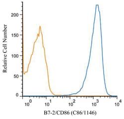
Antigen: B7-2/CD86
Classification: Monoclonal
Concentration: 1.0 mg/mL
Dilution: Western Blot : 0.5 - 1.0 ug/ml, Flow Cytometry : 1 ug/mL, Immunohistochemistry-Paraffin : 2 - 4 ug/ml, Flow (Cell Surface) 1:10-1:1000, Immunofluorescence : 0.5 - 1.0 ug/ml, CyTOF-ready, Knockout Validated
Gene Alias: Activation B7-2 antigen, B70, B7-2, B7-2 antigen, B-lymphocyte activation antigen B7-2, BU63, CD28 antigen ligand 2, CD28LG2B7-2 antigen), CD86 antigen, CD86 molecule, CTLA-4 counter-receptor B7.2, FUN-1, LAB72, MGC34413, T-lymphocyte activation antigen CD86
Host Species: Mouse
Molecular Weight of Antigen: 70 kDa
Quantity: 0.1 mg
Research Discipline: Adaptive Immunity, B Cell Development and Differentiation Markers, Cell Cycle and Replication, Dendritic Cell Markers, Diabetes Research, Immunology, Myeloid derived Suppressor Cell
Gene ID (Entrez): 942
Target Species: Human, Mouse, Rat
Form: Purified
Applications: Western Blot, Flow Cytometry, Immunohistochemistry (Paraffin), Immunofluorescence, CyTOF
Clone: C86/1146
Conjugate: Unconjugated
Formulation: PBS with No Preservative
Gene Symbols: CD86
Immunogen: Recombinant human full-length CD86 protein
Purification Method: Protein A or G purified
Regulatory Status: RUO
Primary or Secondary: Primary
Test Specificity: Recognizes a protein of 70kDa, which is identified as CD86. CD86 is a type I transmembrane glycoprotein and a member of the immunoglobulin superfamily of cell surface receptors. It is expressed at high levels on resting peripheral monocytes and dendritic cells and at very low density on resting B and T lymphocytes. CD86 expression is rapidly upregulated by B cell specific stimuli with peak expression at 18 to 42 hours after stimulation. CD86, along with CD80/B71, is an important accessory molecule in T cell co-stimulation via its interaction with CD28 and CD152/CTLA4. Since CD86 has rapid kinetics of induction, it is believed to be the major CD28 ligand expressed early in the immune response. It is also found on malignant Hodgkin and Reed Sternberg (HRS) cells in Hodgkin's disease.
Content And Storage: Store at 4C short term. Aliquot and store at -20C long term. Avoid freeze-thaw cycles.
Isotype: IgG1 κ
Antigen: B7-2/CD86
Classification: Monoclonal
Concentration: 1.0 mg/mL
Dilution: Western Blot : 0.5 - 1.0 ug/ml, Flow Cytometry : 1 ug/mL, Immunohistochemistry-Paraffin : 2 - 4 ug/ml, Flow (Cell Surface) 1:10-1:1000, Immunofluorescence : 0.5 - 1.0 ug/ml, CyTOF-ready, Knockout Validated
Gene Alias: Activation B7-2 antigen, B70, B7-2, B7-2 antigen, B-lymphocyte activation antigen B7-2, BU63, CD28 antigen ligand 2, CD28LG2B7-2 antigen), CD86 antigen, CD86 molecule, CTLA-4 counter-receptor B7.2, FUN-1, LAB72, MGC34413, T-lymphocyte activation antigen CD86
Host Species: Mouse
Molecular Weight of Antigen: 70 kDa
Quantity: 0.2 mg
Research Discipline: Adaptive Immunity, B Cell Development and Differentiation Markers, Cell Cycle and Replication, Dendritic Cell Markers, Diabetes Research, Immunology, Myeloid derived Suppressor Cell
Gene ID (Entrez): 942
Target Species: Human, Mouse, Rat
Form: Purified
Applications: Western Blot, Flow Cytometry, Immunohistochemistry (Paraffin), Immunofluorescence, CyTOF
Clone: C86/1146
Conjugate: Unconjugated
Formulation: PBS with No Preservative
Gene Symbols: CD86
Immunogen: Recombinant human full-length CD86 protein
Purification Method: Protein A or G purified
Regulatory Status: RUO
Primary or Secondary: Primary
Test Specificity: Recognizes a protein of 70kDa, which is identified as CD86. CD86 is a type I transmembrane glycoprotein and a member of the immunoglobulin superfamily of cell surface receptors. It is expressed at high levels on resting peripheral monocytes and dendritic cells and at very low density on resting B and T lymphocytes. CD86 expression is rapidly upregulated by B cell specific stimuli with peak expression at 18 to 42 hours after stimulation. CD86, along with CD80/B71, is an important accessory molecule in T cell co-stimulation via its interaction with CD28 and CD152/CTLA4. Since CD86 has rapid kinetics of induction, it is believed to be the major CD28 ligand expressed early in the immune response. It is also found on malignant Hodgkin and Reed Sternberg (HRS) cells in Hodgkin's disease.
Content And Storage: Store at 4C short term. Aliquot and store at -20C long term. Avoid freeze-thaw cycles.
Isotype: IgG1 κ