PCNA Antibody (PCNA/694) - IHC-Prediluted, Novus Biologicals™
Manufacturer: Novus Biologicals
Select a Size
| Pack Size | SKU | Availability | Price |
|---|---|---|---|
| Each of 1 | NBP248284-Each-of-1 | In Stock | ₹ 46,636.00 |
NBP248284 - Each of 1
In Stock
Quantity
1
Base Price: ₹ 46,636.00
GST (18%): ₹ 8,394.48
Total Price: ₹ 55,030.48
Antigen
PCNA
Classification
Monoclonal
Conjugate
Unconjugated
Formulation
10mM PBS and 0.05% BSA with 0.05% Sodium Azide
Gene Symbols
PCNA
Immunogen
Recombinant full length human PCNA protein (Uniprot: P12004)
Purification Method
Protein A or G purified
Regulatory Status
RUO
Primary or Secondary
Primary
Test Specificity
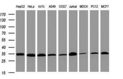
Recognizes a non-histone protein of 36kDa, which is identified as proliferating cell nuclear antigen (PCNA). It is also known as cyclin or polymerase delta auxiliary protein. Elevated expression of PCNA/cyclin has been shown in the nucleus during late G1 phase immediately before the onset of DNA synthesis, becoming maximal during S-phase and declining during G2 and M phases. This monoclonal antibody is excellent for multiple applications.
Content And Storage
Store at 4C.
Isotype
IgG2a κ
Applications
Immunohistochemistry (Paraffin)
Clone
PCNA/694
Dilution
Immunohistochemistry-Paraffin
Gene Alias
cyclin, DNA polymerase delta auxiliary protein, MGC8367, proliferating cell nuclear antigen
Host Species
Mouse
Molecular Weight of Antigen
36 kDa
Quantity
7 mL
Research Discipline
Autophagy, Base Excision Repair, Cell Cycle and Replication, Cellular Markers, Core ESC Like Genes, DNA Polymerases, DNA Repair, Loading Controls, Stem Cell Markers
Gene ID (Entrez)
5111
Target Species
Human
Form
Purified
Related Products
Description
- PCNA Monoclonal specifically detects PCNA in Human samples
- It is validated for Immunohistochemistry, Immunohistochemistry-Paraffin.
Compare Similar Items
Show Difference

Antigen: PCNA
Classification: Monoclonal
Conjugate: Unconjugated
Formulation: 10mM PBS and 0.05% BSA with 0.05% Sodium Azide
Gene Symbols: PCNA
Immunogen: Recombinant full length human PCNA protein (Uniprot: P12004)
Purification Method: Protein A or G purified
Regulatory Status: RUO
Primary or Secondary: Primary
Test Specificity: Recognizes a non-histone protein of 36kDa, which is identified as proliferating cell nuclear antigen (PCNA). It is also known as cyclin or polymerase delta auxiliary protein. Elevated expression of PCNA/cyclin has been shown in the nucleus during late G1 phase immediately before the onset of DNA synthesis, becoming maximal during S-phase and declining during G2 and M phases. This monoclonal antibody is excellent for multiple applications.
Content And Storage: Store at 4C.
Isotype: IgG2a κ
Applications: Immunohistochemistry (Paraffin)
Clone: PCNA/694
Dilution: Immunohistochemistry-Paraffin
Gene Alias: cyclin, DNA polymerase delta auxiliary protein, MGC8367, proliferating cell nuclear antigen
Host Species: Mouse
Molecular Weight of Antigen: 36 kDa
Quantity: 7 mL
Research Discipline: Autophagy, Base Excision Repair, Cell Cycle and Replication, Cellular Markers, Core ESC Like Genes, DNA Polymerases, DNA Repair, Loading Controls, Stem Cell Markers
Gene ID (Entrez): 5111
Target Species: Human
Form: Purified
Antigen: PD-1
Classification: Monoclonal
Conjugate: Unconjugated
Formulation: 10mM PBS and 0.05% BSA with 0.05% Sodium Azide
Gene Symbols: PDCD1
Immunogen: Recombinant full-length human PD-1 protein (Uniprot: Q15116)
Purification Method: Protein A or G purified
Regulatory Status: RUO
Primary or Secondary: Primary
Test Specificity: PDCD-1 (programmed cell death-1 protein), also designated CD279, is a type I transmembrane receptor and a member of the immunoglobin gene superfamily. It is expressed on activated T-cells, B-cells, and myeloid cells. Anti-PDCD-1 is a marker of angioimmunoblastic lymphoma and suggests a unique cell of origin for this neoplasm. Unlike CD10 and BCL6, PDCD-1 is expressed by few B-cells, so anti-PDCD-1 may be a more specific and useful diagnostic marker in angioimmunoblastic lymphoma. In addition, PDCD-1 expression provides evidence that angioimmunoblastic lymphoma is a neoplasm derived from germinal center-associated T-cells.
Content And Storage: Store at 4C.
Isotype: IgG1 κ
Applications: Immunohistochemistry (Paraffin)
Clone: PDCD1/922
Dilution: Immunohistochemistry-Paraffin
Gene Alias: CD279, CD279 antigen, hPD-1, PD1hPD-l, programmed cell death 1, programmed cell death protein 1, Protein PD-1, SLEB2
Host Species: Mouse
Molecular Weight of Antigen: 55 kDa
Quantity: 7 mL
Research Discipline: Apoptosis, Stem Cell Markers
Gene ID (Entrez): 5133
Target Species: Human
Form: Purified
Antigen: PD-1
Classification: Monoclonal
Conjugate: Unconjugated
Formulation: 10mM PBS and 0.05% BSA with 0.05% Sodium Azide
Gene Symbols: PDCD1
Immunogen: Recombinant full-length human PD-1 protein (Uniprot: Q15116)
Purification Method: Protein A or G purified
Regulatory Status: RUO
Primary or Secondary: Primary
Test Specificity: PDCD-1 (programmed cell death-1 protein), also designated CD279, is a type I transmembrane receptor and a member of the immunoglobin gene superfamily. It is expressed on activated T-cells, B-cells, and myeloid cells. Anti-PDCD-1 is a marker of angioimmunoblastic lymphoma and suggests a unique cell of origin for this neoplasm. Unlike CD10 and BCL6, PDCD-1 is expressed by few B-cells, so anti-PDCD-1 may be a more specific and useful diagnostic marker in angioimmunoblastic lymphoma. In addition, PDCD-1 expression provides evidence that angioimmunoblastic lymphoma is a neoplasm derived from germinal center-associated T-cells.
Content And Storage: Store at 4C.
Isotype: IgG1 κ
Applications: Immunohistochemistry (Paraffin)
Clone: SPM597
Dilution: Immunohistochemistry-Paraffin
Gene Alias: CD279, CD279 antigen, hPD-1, PD1hPD-l, programmed cell death 1, programmed cell death protein 1, Protein PD-1, SLEB2
Host Species: Mouse
Molecular Weight of Antigen: 55 kDa
Quantity: 7 mL
Research Discipline: Apoptosis, Stem Cell Markers
Gene ID (Entrez): 5133
Target Species: Human
Form: Purified