HLA DQ/DR/DP Antibody (CR3/43), DyLight 488, Novus Biologicals™
Manufacturer: Novus Biologicals
Select a Size
| Pack Size | SKU | Availability | Price |
|---|---|---|---|
| Each of 1 | NBP254507G-Each-of-1 | In Stock | ₹ 56,426.00 |
NBP254507G - Each of 1
In Stock
Quantity
1
Base Price: ₹ 56,426.00
GST (18%): ₹ 10,156.68
Total Price: ₹ 66,582.68
Antigen
HLA DPB1
Classification
Monoclonal
Conjugate
DyLight 488
Formulation
50 mM sodium borate with 0.05% sodium azide
Gene Symbols
HLA-DPB1
Immunogen
Cells from human tonsil
Primary or Secondary
Primary
Test Specificity
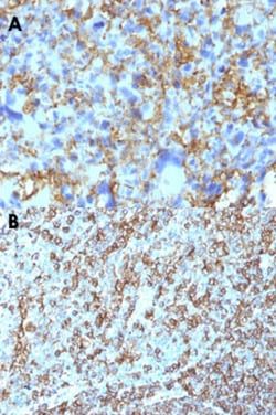
Reacts with a common epitope of human major histocompatibility (MHC) class II antigens, HLA-DP, -DQ and -DR. Human MHC class II antigens are transmembrane glycoproteins composed of an chain (36kDa) and a chain (27kDa). They are expressed primarily on antigen presenting cells such as B lymphocytes, monocytes, macrophages, and thymic epithelial cells and are also present on activated T lymphocytes. Human MHC class II genes are located in the HLA-D region that encodes at least six and ten chain genes. Three loci, DR, DQ and DP, encode the major expressed products of the human class II region. The human MHC class II molecules bind intracellularly processed peptides and present them to T-helper cells. They, therefore, have a critical role in the initiation of the immune response. It has been shown that some autoimmune diseases are associated with certain class II alleles.
Content And Storage
Store at 4°C in the dark.
Applications
Western Blot, Flow Cytometry, Immunohistochemistry, Immunocytochemistry, Immunofluorescence, Immunohistochemistry (Paraffin)
Clone
CR3/43
Dilution
Western Blot, Flow Cytometry, Immunohistochemistry, Immunocytochemistry/Immunofluorescence, Immunohistochemistry-Paraffin
Gene Alias
DP beta 1 chain, DPB1, HLA class II histocompatibility antigen, DP(W4) beta chain, HLA DP14-beta chain, HLA-DP histocompatibility type, beta-1 subunit, HLA-DP1B, HLA-DPB, major histocompatibility complex class II HLA DPB1 protein, major histocompatibility complex, class II, DP beta 1, MHC class II antigen beta chain, MHC class II antigen DP beta 1 chain, MHC class II antigen DPB1, MHC class II antigen DPbeta1, MHC class II HLA-DP-beta, MHC HLA DPB1
Host Species
Mouse
Quantity
0.1 mL
Gene ID (Entrez)
3115
Target Species
Human
Isotype
IgG1 κ
Related Products


Description
- HLA DPB1 Monoclonal specifically detects HLA DPB1 in Human samples
- It is validated for Western Blot, Flow Cytometry, ELISA, Immunohistochemistry, Immunocytochemistry/Immunofluorescence, Immunohistochemistry-Paraffin.
Compare Similar Items
Show Difference
Antigen: HLA DPB1
Classification: Monoclonal
Conjugate: DyLight 488
Formulation: 50 mM sodium borate with 0.05% sodium azide
Gene Symbols: HLA-DPB1
Immunogen: Cells from human tonsil
Primary or Secondary: Primary
Test Specificity: Reacts with a common epitope of human major histocompatibility (MHC) class II antigens, HLA-DP, -DQ and -DR. Human MHC class II antigens are transmembrane glycoproteins composed of an chain (36kDa) and a chain (27kDa). They are expressed primarily on antigen presenting cells such as B lymphocytes, monocytes, macrophages, and thymic epithelial cells and are also present on activated T lymphocytes. Human MHC class II genes are located in the HLA-D region that encodes at least six and ten chain genes. Three loci, DR, DQ and DP, encode the major expressed products of the human class II region. The human MHC class II molecules bind intracellularly processed peptides and present them to T-helper cells. They, therefore, have a critical role in the initiation of the immune response. It has been shown that some autoimmune diseases are associated with certain class II alleles.
Content And Storage: Store at 4°C in the dark.
Applications: Western Blot, Flow Cytometry, Immunohistochemistry, Immunocytochemistry, Immunofluorescence, Immunohistochemistry (Paraffin)
Clone: CR3/43
Dilution: Western Blot, Flow Cytometry, Immunohistochemistry, Immunocytochemistry/Immunofluorescence, Immunohistochemistry-Paraffin
Gene Alias: DP beta 1 chain, DPB1, HLA class II histocompatibility antigen, DP(W4) beta chain, HLA DP14-beta chain, HLA-DP histocompatibility type, beta-1 subunit, HLA-DP1B, HLA-DPB, major histocompatibility complex class II HLA DPB1 protein, major histocompatibility complex, class II, DP beta 1, MHC class II antigen beta chain, MHC class II antigen DP beta 1 chain, MHC class II antigen DPB1, MHC class II antigen DPbeta1, MHC class II HLA-DP-beta, MHC HLA DPB1
Host Species: Mouse
Quantity: 0.1 mL
Gene ID (Entrez): 3115
Target Species: Human
Isotype: IgG1 κ
Antigen: Kappa Light Chain
Classification: Monoclonal
Conjugate: DyLight 488
Formulation: 50 mM sodium borate with 0.05% sodium azide
Gene Symbols: IGKC
Immunogen: Recombinant human full-length Ig kappa chain
Primary or Secondary: Primary
Test Specificity: This monoclonal antibody is specific to kappa light chain of immunoglobulin and shows no cross-reaction with lambda light chain or any of the five heavy chains. It recognizes human Ig kappa light chains of both secreted and cell surface immunoglobulin. It detects also free kappa light chains. In mammals, the two light chains in an antibody are always identical, with only one type of light chain, kappa or lambda. The ratio of Kappa to Lambda is 70:30. However, with the occurrence of multiple myeloma or other B-cell malignancies this ratio is disturbed. Antibody to the kappa light chain is reportedly useful in the identification of leukemias, plasmacytomas, and certain non-Hodgkins lymphomas. Demonstration of clonality in lymphoid infiltrates indicates that the infiltrate is malignant.
Content And Storage: Store at 4°C in the dark.
Applications: Flow Cytometry, Immunohistochemistry, Immunohistochemistry (Paraffin)
Clone: KLC1278
Dilution: Flow Cytometry, Immunohistochemistry, Immunohistochemistry-Paraffin
Gene Alias: HCAK1, IGKCD, immunoglobulin kappa constant, Km, MGC111575, MGC62011, MGC72072, MGC88770, MGC88771, MGC88809
Host Species: Mouse
Quantity: 0.1 mL
Gene ID (Entrez): 3514
Target Species: Human
Isotype: IgG1 κ
Antigen: Desmocollin-2
Classification: Monoclonal
Conjugate: DyLight 488
Formulation: 50 mM sodium borate with 0.05% sodium azide
Gene Symbols: DSC2
Immunogen: Human desmocollin-2 extracellular domain (exact sequence is proprietary)
Primary or Secondary: Primary
Test Specificity: __
Content And Storage: Store at 4°C in the dark.
Applications: Western Blot, Flow Cytometry, Immunohistochemistry, Immunocytochemistry, Immunofluorescence, Immunohistochemistry (Paraffin)
Clone: 7G6
Dilution: Western Blot, Flow Cytometry, Immunohistochemistry, Immunocytochemistry/Immunofluorescence, Immunohistochemistry-Paraffin
Gene Alias: ARVD11, Cadherin family member 2, CDHF2DGII/III, desmocollin 2, desmocollin-2, Desmocollin-3, Desmosomal glycoprotein II, desmosomal glycoprotein II/III, Desmosomal glycoprotein III, DG2, DSC3DKFZp686I11137
Host Species: Mouse
Quantity: 0.1 mL
Gene ID (Entrez): 1824
Target Species: Human, Mouse, Rat
Isotype: IgG1 κ